. 2023 Jun 23;9(1):22.
doi: 10.1038/s41405-023-00151-x.

Efficacy of Nano Silver Fluoride and/or Diode Laser In Enhancing Enamel Anticariogenicity around orthodontic brackets

Affiliations

Efficacy of Nano Silver Fluoride and/or Diode Laser In Enhancing Enamel Anticariogenicity around orthodontic brackets

Aya Anwar Alsherif et al. BDJ Open. .

Abstract

Purpose: This in vitro study aimed to compare the anticariogenic effect of using diode laser irradiation and/or nano silver fluoride varnish around orthodontic brackets.

Materials and methods: 60 caries-free and intact premolars were randomly divided into 3 experimental groups as follow: (1) Group I (nano silver fluoride treated group, n = 20), (2) Group II (diode laser treated group, n = 20) and (3) Group III (combined nano silver fluoride and diode laser treated group, n = 20). Anticariogenicity was assessed using polarized light, scanning electron microscope, elemental and shear bond strength analyses.

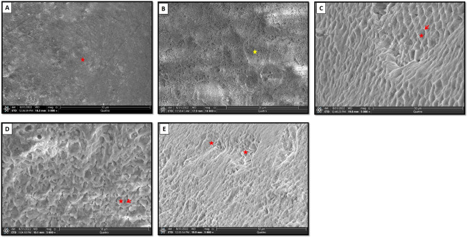
Results: PLM and SEM showed presence of few demineralized areas in group I. Group II revealed a dramatic increased demineralization. Group III disclosed almost typical homogenous surface enamel. elemental analysis showed a highly significant difference between Group III and II and a significant difference between Group III and I. Shear bond strength analysis revealed a significant difference between group I and II and between group III and II. The difference between group III and I was non-significant.

Conclusion: Both diode laser and nano silver fluoride positively affected dental enamel with the most superior enhancement in enamel criteria was achieved by surface pretreatment by combined nano silver fluoride varnish and diode laser irradiation.


Conflict of interest statement

The authors declare no competing interests.

Figures

Fig. 1
Fig. 1. Flow chart illustrating the study design.
(Inset) Photomicrograph showing sectioning of the buccal surface into right side (test side) and left side (control side).
Fig. 2
Fig. 2. PLM of group I-B, group I-A, group II-A and group III-A.
Polarized light photomicrographs of longitudinal ground sections showing, (A): group I section-B displaying a strong positive birefringent demineralized enamel band (double head arrow) extending beneath an intact surface layer (red arrows). B Group I section-A showing normal subsurface enamel (double head arrow) with few demineralized areas (black arrow). C Group II section-A exhibiting a widely distributed demineralized areas (double arrow head), though, still displaying minimal areas of remineralized enamel (black arrows). D Group III section-A showing almost typical homogenous subsurface enamel (double head arrow), reflecting normal mineralization and birefringence of enamel. (PLM, original magnification; A, B, C, D × 40).
Fig. 3
Fig. 3. Scanning electron micrographs of normal enamel specimen and section B of all groups.
A Showing smooth enamel architecture with homogenous aprismatic layer covering its surface (asterisk). B A higher magnification showing typical fish scale appearance (asterisk). C Showing type 1etching pattern with preferential removal of rod core (asterisk). Arrow; intact rod periphery. D Showing type II etching pattern where rod periphery was selectively removed (arrow). Asterisk; rod core. E Showing irregular etching pattern (asterisk). (SEM, original magnification; A, CE × 5000, B × 20,000).
Fig. 4
Fig. 4. Scanning electron micrographs of group I section A, Group II section A and Group III section A.
A, B Group I-A showing calcium deposits along the majority of enamel surface (red arrows) with detection of few micropores (black arrow). Residual NSF particles were adhered to enamel surface (yellow arrows). C Group II-A showing micro-cracks (red arrows) with detection of many scattered micro-pores (yellow arrows). D A higher magnification showing uneven enamel crystals with many porosities (yellow arrows) and minor amorphous surface precipitation (red arrows). E Group II-A showing homogenous enamel surface layer (asterisk). F A higher magnification of the boxed area of (E) showing minimal micro-pores (boxed area). Asterisk; homogenous enamel surface. (SEM, original magnification; A, C, E × 5000, B, D, F × 20,000).

References

    1. Tufekci E, Dixon JS, Gunsolley JC, Lindauer SJ. Prevalence of white spot lesions during orthodontic treatment with fixed appliances. Angle Orthod. 2011;81:10–4. doi: 10.2319/051710-262.1. - DOI - PMC - PubMed
    1. Liu H, Sun J, Dong Y, Lu H, Zhou H, Hansen BF, et al. Periodontal health and relative quantity of subgingival Porphyromonas gingivalis during orthodontic treatment. Angle Orthod. 2011;81:609–15. doi: 10.2319/082310-352.1. - DOI - PMC - PubMed
    1. Lucchese A, Gherlone E. Prevalence of white-spot lesions before and during orthodontic treatment with fixed appliances. Eur J Orthod. 2013;35:664–8. doi: 10.1093/ejo/cjs070. - DOI - PubMed
    1. Bishara SE, Ostby AW. White spot lesions: formation, prevention, and treatment. in Seminars in orthodontics, Vol. 14. Elsevier; 2008. p. 174–82.
    1. Al Maaitah EF, Adeyemi AA, Higham SM, Pender N, Harrison JE. Factors affecting demineralization during orthodontic treatment: a post-hoc analysis of RCT recruits. Am J Orthod Dentofac Orthop. 2011;139:181–91. doi: 10.1016/j.ajodo.2009.08.028. - DOI - PubMed