. 2023 Jun 23;14(1):3760.
doi: 10.1038/s41467-023-39032-0.

Rapid and reversible optical switching of cell membrane area by an amphiphilic azobenzene

Affiliations

Rapid and reversible optical switching of cell membrane area by an amphiphilic azobenzene

Fabian Höglsperger et al. Nat Commun. .

Abstract

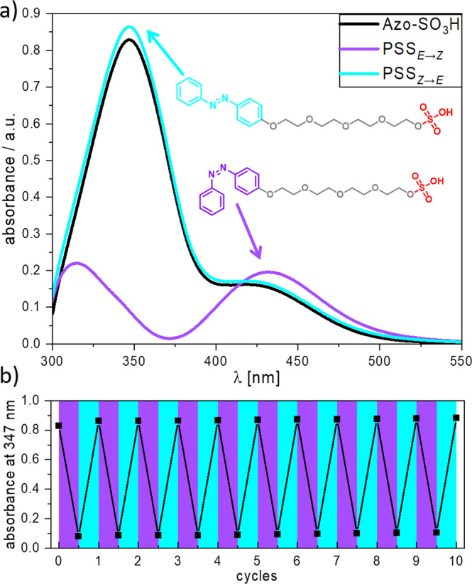
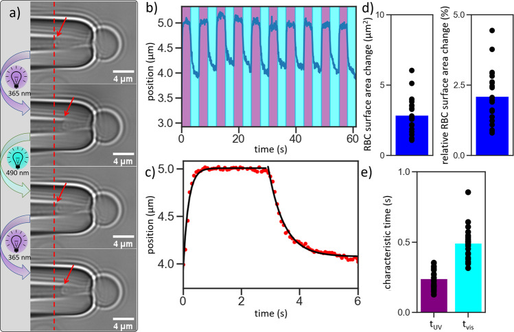
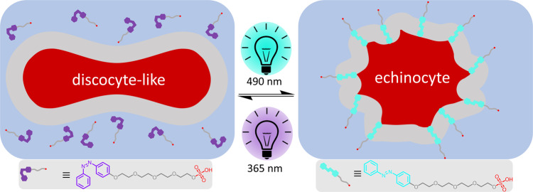
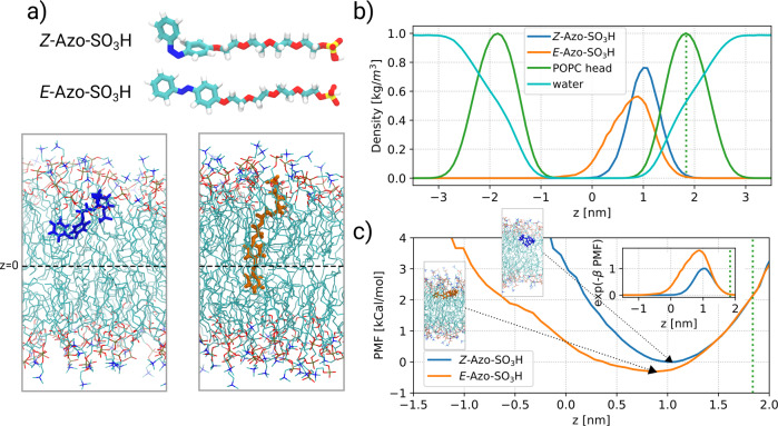
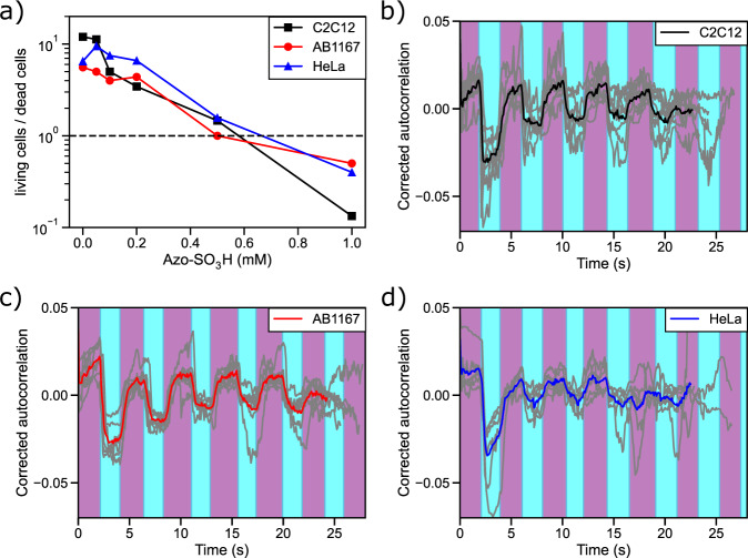
Cellular membrane area is a key parameter for any living cell that is tightly regulated to avoid membrane damage. Changes in area-to-volume ratio are known to be critical for cell shape, but are mostly investigated by changing the cell volume via osmotic shocks. In turn, many important questions relating to cellular shape, membrane tension homeostasis and local membrane area cannot be easily addressed because experimental tools for controlled modulation of cell membrane area are lacking. Here we show that photoswitching an amphiphilic azobenzene can trigger its intercalation into the plasma membrane of various mammalian cells ranging from erythrocytes to myoblasts and cancer cells. The photoisomerization leads to a rapid (250-500 ms) and highly reversible membrane area change (ca 2 % for erythrocytes) that triggers a dramatic shape modulation of living cells.


Conflict of interest statement

The authors declare no competing interests.

Figures

Fig. 1
Fig. 1. Reversible photoisomerization of Azo-SO3H.
a Molecular structure of the E- and Z-isomer of photoswitch Azo-SO3H and UV/vis absorption spectra in the initial state and the two photostationary states PSSE→Z after 5 s UV (365 nm) irradiation and PSSZ→E after 10 s blue (465 nm) irradiation (80 µM in DMEM). b Photoswitching cycles (n = 10) for Azo-SO3H using alternating UV (5 s at 356 nm) and blue irradiation (10 s at 465 nm).
Fig. 2
Fig. 2. Microscopy images of free-floating RBCs.
ad The morphology changes depend on the reversible photoisomerization of the Azo-SO3H from E-Isomer (echinocyte, (a) and (c)) to Z-Isomer (discocyte-like, (b) and (d)). e Quantification of the inhomogeneity of N = 3, n = 13 RBCs via the standard deviation of the pixel values. The difference between the discocyte-like and echinocyte is significant (two-sided t test, p = 7 × 10⁻⁷).
Fig. 3
Fig. 3. Analysis of aspirated RBCs with low applied tension of ca.
10-5 N/m. a Microscopy images from an aspirated RBC during photoisomerization with consequent movement of the membrane (red line and arrow as a guide). b Tracking of the movement of the membrane inside the micropipette while switching for 10 cycles. c Time-dependent movement of 10 cycle shifted on top of each other, averaged and fitted (black line). d Analysis of the absolute change in membrane area and in relation to the overall surface. Data show 22 experiments on 6 different days. e Characteristic switching times, tUV: UV light switch, tvis: vis light switch. Data show N = 6, n = 22 experiments.
Fig. 4
Fig. 4. Relaxation times of aspirated RBCs depends on the light intensity.
a Relaxation after vis light irradiation. b Relaxation after UV light irradiation. The black dashed line is the average relaxation time after irradiation with 100% vis or 2% UV intensity, respectively (see Fig. 3e). For light intensity translation from % to µW see Supplementary Fig. 8. The red dashed line is an exponential function of the fitted characteristic switching times with increasing irradiation intensity and serves as a guide to the eye, while the gray curves represent individual measurements. Data show N = 3, n = 7 (a) and N = 3, n = 8 (b) experiments.
Fig. 5
Fig. 5. Photoresponsive shape transformation of RBCs.
Schematic representation of the photoresponsive, reversible incorporation of Azo-SO3H into the RBC plasma membrane and the corresponding switch between the discocyte-like and echinocyte morphology.
Fig. 6
Fig. 6. Molecular dynamics simulation of Azo-SO3H a lipid membrane.
a Molecular structure of the Azo-SO3H isomers and snapshots of the E-isomer and Z-isomer inside the POPC membrane. The approximate position of the center of the membrane (z = 0) is shown by a dashed line. b Number densities of Azo-SO3H as well as the POPC head group and the water molecules along the membrane normal (z-axis). The number densities for the POPC head group and water molecules were rescaled and multiplied by 0.017 and 0.01, respectively, so that all the profiles can be conveniently shown in one plot. The average value of the density profile of the POPC head group is shown by a dotted line. c The PMF profiles of the Azo-SO3H molecules along the membrane normal are shown. The snapshot of the two isomers for the minimum of the free energy is represented. The PMF-profiles are shifted so that they agree at the average position of the POPC head group. The resulting distribution exp(-β PMF) for the two isomers is shown in the inset.
Fig. 7
Fig. 7. Photomanipulation of myoblasts and cancer cells with Azo-SO3H.
Viability assay of the three different cell lines (a) and the corrected autocorrelation profiles that quantify the changes in cell morphology over 5 cycles at concentrations of 0.5 mM Azo-SO3H (bd) on C2C12 (N = 3, n = 8, b), AB1167 (N = 3, n = 7, c) and HeLa cells (N = 3, n = 6, d).

References

    1. McMahon HT, Gallop JL. Membrane curvature and mechanisms of dynamic cell membrane remodelling. Nature. 2005;438:590–596. doi: 10.1038/nature04396. - DOI - PubMed
    1. Benmerah A, Lamaze C. Clathrin-coated pits: vive la différence? Traffic. 2007;8:970–982. doi: 10.1111/j.1600-0854.2007.00585.x. - DOI - PubMed
    1. Parton RG, Kozlov MM, Ariotti N. Caveolae and lipid sorting: shaping the cellular response to stress. J. Cell. Biol. 2020;219:e201905071. doi: 10.1083/jcb.201905071. - DOI - PMC - PubMed
    1. Sackmann, E., Reuther, A. & Heinrich, D. Micromechanics of cells. In Mechanics of the 21st century. Proc. 21st International Congress of Theoretical and Applied Mechanics, Warsaw, Poland, 15–21 August 2004 (ed Gutkowski, W. & Kowalewski, T. A.) 313–328 (Springer, 2005).
    1. Belly Hde, et al. Membrane tension gates ERK-mediated regulation of pluripotent cell fate. Cell Stem Cell. 2021;28:273–284.e6. doi: 10.1016/j.stem.2020.10.018. - DOI - PMC - PubMed

Publication types