. 2023 Jun 23;14(1):3749.
doi: 10.1038/s41467-023-39336-1.

A joint proteomic and genomic investigation provides insights into the mechanism of calcification in coccolithophores

Affiliations

A joint proteomic and genomic investigation provides insights into the mechanism of calcification in coccolithophores

Alastair Skeffington et al. Nat Commun. .

Abstract

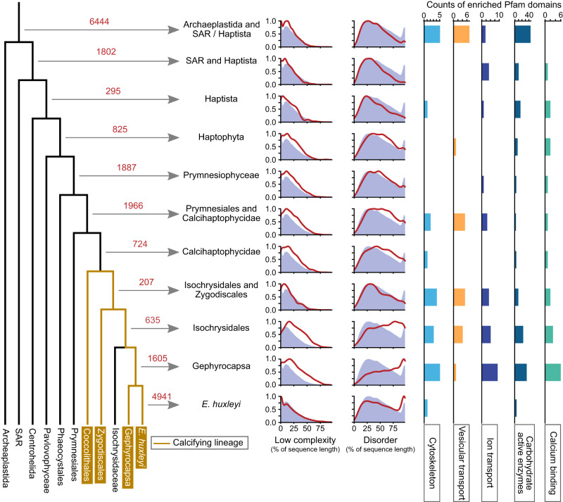
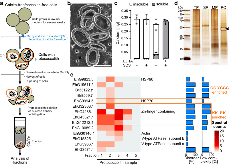
Coccolithophores are globally abundant, calcifying microalgae that have profound effects on marine biogeochemical cycles, the climate, and life in the oceans. They are characterized by a cell wall of CaCO3 scales called coccoliths, which may contribute to their ecological success. The intricate morphologies of coccoliths are of interest for biomimetic materials synthesis. Despite the global impact of coccolithophore calcification, we know little about the molecular machinery underpinning coccolithophore biology. Working on the model Emiliania huxleyi, a globally distributed bloom-former, we deploy a range of proteomic strategies to identify coccolithogenesis-related proteins. These analyses are supported by a new genome, with gene models derived from long-read transcriptome sequencing, which revealed many novel proteins specific to the calcifying haptophytes. Our experiments provide insights into proteins involved in various aspects of coccolithogenesis. Our improved genome, complemented with transcriptomic and proteomic data, constitutes a new resource for investigating fundamental aspects of coccolithophore biology.


Conflict of interest statement

The authors declare no competing interests.

Figures

Fig. 1
Fig. 1. Properties of the E. huxleyi genome and proteome v2.
a Numbers of isoforms detected in genes derived from the Isoseq data. b Comparison of Emihu1 predicted best gene models and the gene models derived from the Isoseq data using GFFCompare. Emihu1 transcripts were either compared to Isoseq data through mapping to the Emihu2 genome or Isoseq transcripts were compared to Emihu1 transcripts through mapping to the Emihu1 genome. c Comparison of the new predicted proteome (from Isoseq derived, plus de novo predicted gene models) to the Emihu1 best predicted proteome using DIAMOND blastp. Proteins were categorized according to the e-value (eval.) of the top High-scoring Segment Pair, requiring a query coverage of either 100% or >50%. d Comparison of identification rates of spectra clusters (where only high-quality spectra were used, and each cluster likely represents a unique peptide) using the new E. huxleyi protein database Emihu2, and the Emihu1 best proteins database. Rates are shown for four independent E. huxleyi whole-cell detergent extracts (1–4) and for three independent extracts derived from isolated coccoliths (5–7).
Fig. 2
Fig. 2. Phylostratigraphic analysis of new E. huxleyi proteome.
Orthogroups were calculated for predicted proteomes of 38 species, including E. huxleyi, covering the phylogenetic range shown in the schematic tree. Red numbers indicate the number of orthogroups containing E. huxleyi sequences, that arise at each branch of the tree. For example, 825 orthogroups with E. huxleyi sequences arose in the common ancestor of all haptophytes. For each group, density plots of the proportion of each protein sequence that is low complexity and the proportion that is disordered are shown as red lines, while the purple background indicates the distribution for the entire E. huxleyi proteome. Pfam domains enriched in each orthogroups relative to the entire proteome were collated into categories and the counts of enriched domains for each category is displayed.
Fig. 3
Fig. 3. CAP biosynthesis is maintained during low calcium growth.
a Experimental design for probing CAP biosynthesis in low calcium grown cells. Cells grown in low calcium medium (low-Ca), in which they do not produce calcite, were aliquoted into two cultures. To one aliquot, calcium was added to induce calcite formation (std-Ca), while the other aliquot was continued in a low-calcium medium. At 0, 1, 3, and 6 h after calcium addition, samples were taken for microscopic and proteomics analyses. Extracellular polysaccharides were isolated from stationary phase cultures, as described in the materials and methods. The low-Ca culture for polysaccharide isolation was started with cells that had been propagated in a low-Ca medium for 1.5 months. The recalcification experiment was performed with cells that had been in a low-Ca medium for 14 days. b Monosaccharide composition of the acid-hydrolyzed extracellular polysaccharide samples of low-Ca and std-Ca grown cultures and CAP extract from isolated coccoliths expressed as molar ratio as determined by HPAEC-PAD analysis. The numbers give the molar ratio of each monosaccharide to mannose, which was the most abundant monosaccharide in the main CAP of E. huxleyi. Note that not all monosaccharides that were detected could be quantified. For chromatograms see Supplementary Fig. 6. c SDS-PAGE analysis (n = 3, n = biologically independent samples) of extracellular polysaccharide samples and CAP extract stained with the cationic dye Stains-all. Arrowhead marks the dominant CAP of E. huxleyi coccoliths. Source data are provided as Source Data file.
Fig. 4
Fig. 4. Identification of coccolith proteins.
a Silver-stained Tricine-SDS-PAGE (n = 3, n = biologically independent samples) of soluble coccolith-associated organic material (SCAOM) isolated from purified coccoliths (Supplementary Fig. 7) and chemically deglycosylated SCAOM without further treatment (−Trypsin) and after treatment with trypsin (+Trypsin). The arrowhead marks the dominant CAP of E. huxleyi. Note that unlike Stains-all staining (Fig. 3c), the CAP appears as a negative band in a silver-stained gel. b Schematic primary structures of the COPROs.
Fig. 5
Fig. 5. Enrichment and proteomics analysis of proto-coccoliths.
a Experimental setup for the enrichment of proto-coccoliths. Cells were ruptured in a French press and aggregates and unbroken cells were removed by low-speed centrifugation. Proto-coccoliths were enriched by two consecutive rounds of sucrose density centrifugation. The last five fractions from the bottom were proteomically analyzed. b SEM micrograph of the material in the bottom fraction, showing that it contains proto-coccoliths. (n = 2, n = independent biological replicates). c Fractionation of calcium in suspensions of proto-coccoliths treated with (+) and without (−) SDS and EDTA, determined by ICP-OES. Isolated proto-coccolith calcite is protected from dissolution by the calcium chelator EDTA. After the addition of SDS detergent, which solubilizes membranes, proto-coccolith calcite is dissolved by EDTA. Data are represented as mean ± SD (n = 3, n = independent biological replicates). d Silver-stained SDS-PAGE gel of protein extracts (n = 3, n = independent biological replicates) from whole-cells (TP), soluble proteins (SP), membrane-bound proteins (MP), and enriched proto-coccoliths (PC). The arrowhead marks the main CAP of E. huxleyi, which associates with the proto-coccolith calcite in the CV. e Distribution of proteins across the bottom fractions of a proto-coccolith. For each protein, the percentage of the sequence that is disordered and the percentage that is of low complexity is given. Proteins enriched in specific motifs are boxed.
Fig. 6
Fig. 6. The proteome of the coccosphere.
a Schematics of a selection of putative coccosphere proteins that may have a role in calcification, identified in at least three replicates and with conserved domains. b Schematics of putative coccosphere proteins without conserved domains, also identified among our other datasets (see below for CvsN dataset). Overlaps with other datasets are indicated (CvsN C-cells vs N-cells dataset, LvsD Light vs Dark dataset, CV coccolith vesicle, COPRO coccolith protein) as are sequence motifs enriched in the coccosphere proteins relative to the entire proteome.
Fig. 7
Fig. 7. Overlap between datasets.
‘C-cells vs N-cells’ and ‘Light vs Dark’ datasets are proteins regulated in either direction, q-value < 0.05. Coccosphere proteins are Std-Ca-specific proteins identified in at least two replicates by at least two peptides. COPROs are as described above. CV proteins are those found exclusively in the C-cell density gradients.
Fig. 8
Fig. 8. Model for the involvement of proteins in coccolith formation.
Proposed molecular organization of the CV and the coccosphere. At standard calcium concentration (Std-Ca), coccoliths form inside the specialized CV, where calcite nucleation and morphogenesis are controlled by luminal polysaccharides (CAPs, light blue) and proteins, as well as cytosolic proteins interacting with the CV membrane (orange). The inset (top left) shows the hypothesized distribution of selected proteins identified in two or more proteomic experiments inside and outside the CV and their proposed function. An intriguing candidate CV luminal protein is EhG42278.1 which possesses multiple Ser-(Pro)3-4 peptide repeats, defining structural features of glycosylated algal and plant cell wall proteins known as extensins, which play a crucial role in cell wall assembly. EhG42278.1 may play a comparable role to the extensins in the assembly of the base plates. Other strong candidates for CV luminal proteins are the double pentapeptide motif-containing proteins EhG21037.1 and EhG40669.2, EhG33272.1, and EhG6475.1. V-type ATPase in the CV membrane may regulate the pH in the CV lumen, and the cytosolic proteins actin and myosin may be involved in CV morphogenesis and coccolith extrusion. The coccosphere (inset bottom left) is an environment rich in proteins, some of which, such as carbonic anhydrase, may have roles in coccolith biogenesis. Most of the coccosphere candidate proteins (17 in total, 16 globular and one transmembrane protein) are of unknown function. The exchange of ions between the cell and the environment is facilitated by a plethora of ion transporters and channels, of which five were identified here (HCO3 transporter: EhG9529.1, Anion channel: EhG33980.1, Na+/Ca2+ K+ exchanger: EhG40126.1, Mg2+ transporter: EhG42190.1, ABC transporter: EhG35076.1). At low calcium concentrations (right side), calcite formation ceases, but not CAP biosynthesis and secretion, and therefore we propose that extracellular calcium availability is not an activator of the genetic program underlying coccolith formation.

References

    1. Balch WM. The ecology, biogeochemistry, and optical properties of coccolithophores. Ann. Rev. Mar. Sci. 2018;10:71–98. doi: 10.1146/annurev-marine-121916-063319. - DOI - PubMed
    1. Alcolombri U, et al. Identification of the algal dimethyl sulfide-releasing enzyme: a missing link in the marine sulfur cycle. Science. 2015;348:1466–1469. doi: 10.1126/science.aab1586. - DOI - PubMed
    1. Segev E, et al. Dynamic metabolic exchange governs a marine algal-bacterial interaction. Elife. 2016;5:e17473. doi: 10.7554/eLife.17473. - DOI - PMC - PubMed
    1. Schatz D, et al. Communication via extracellular vesicles enhances viral infection of a cosmopolitan alga. Nat. Microbiol. 2017;2:1485–1492. doi: 10.1038/s41564-017-0024-3. - DOI - PubMed
    1. Schatz D, et al. Ecological significance of extracellular vesicles in modulating host-virus interactions during algal blooms. ISME J. 2021;15:3714–3721. doi: 10.1038/s41396-021-01018-5. - DOI - PMC - PubMed

Publication types