. 2023 Jun 23;22(1):99.
doi: 10.1186/s12943-023-01791-1.

m6A methylation reader IGF2BP2 activates endothelial cells to promote angiogenesis and metastasis of lung adenocarcinoma

Affiliations

m6A methylation reader IGF2BP2 activates endothelial cells to promote angiogenesis and metastasis of lung adenocarcinoma

Han Fang et al. Mol Cancer. .

Abstract

Background: Lung adenocarcinoma (LUAD) is a common type of lung cancer with a high risk of metastasis, but the exact molecular mechanisms of metastasis are not yet understood.

Methods: This study acquired single-cell transcriptomics profiling of 11 distal normal lung tissues, 11 primary LUAD tissues, and 4 metastatic LUAD tissues from the GSE131907 dataset. The lung multicellular ecosystems were characterized at a single-cell resolution, and the potential mechanisms underlying angiogenesis and metastasis of LUAD were explored.

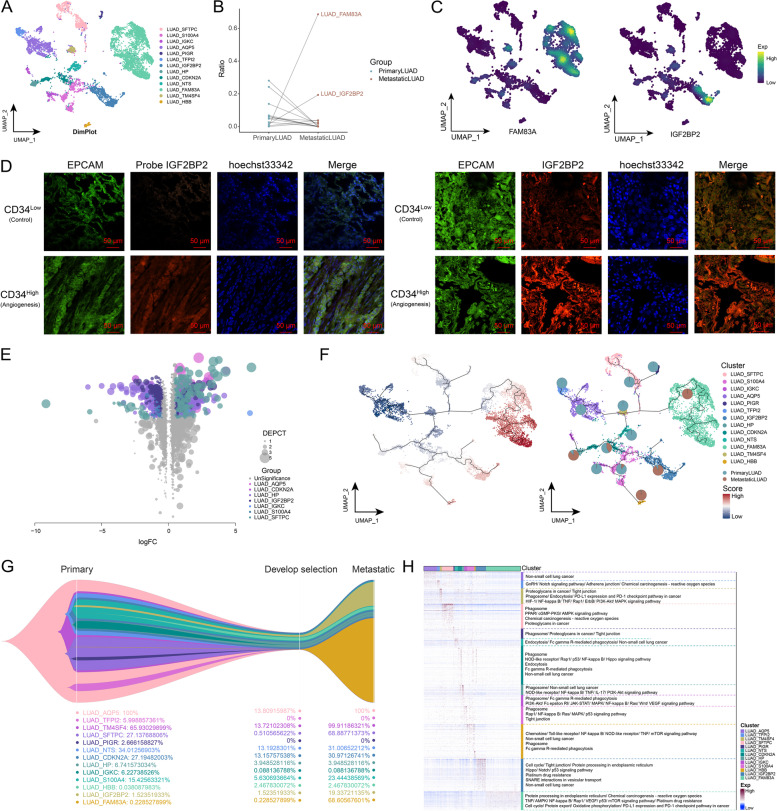
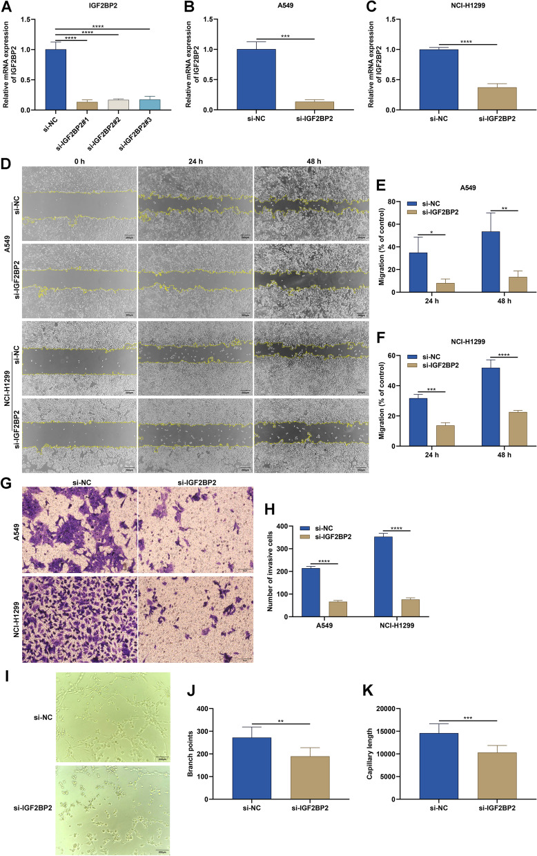
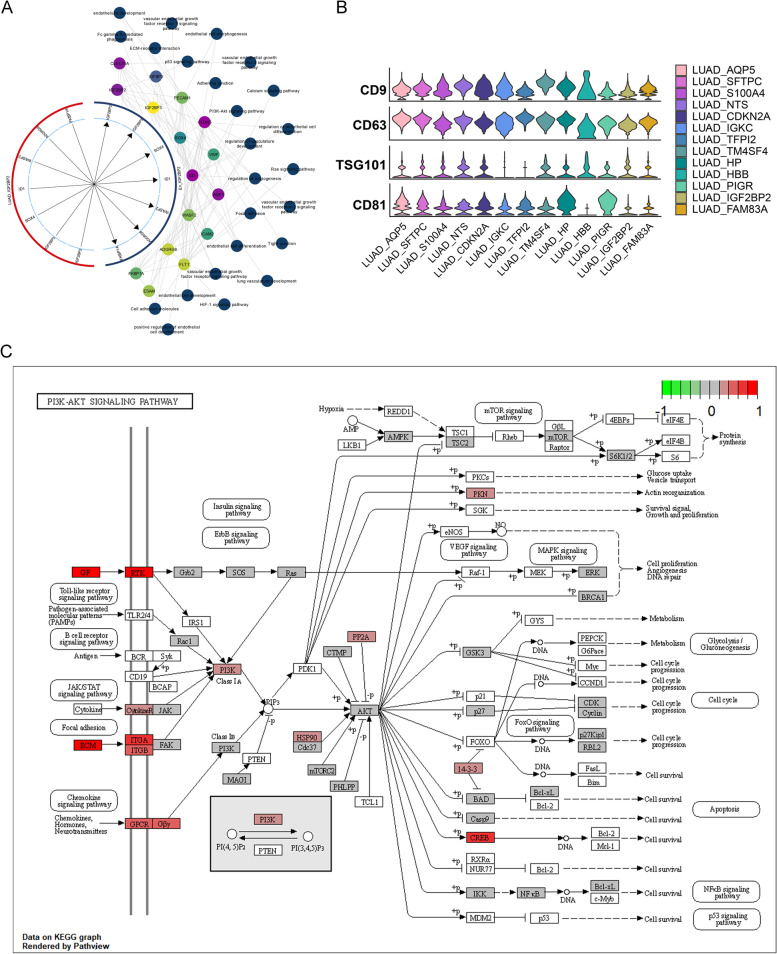
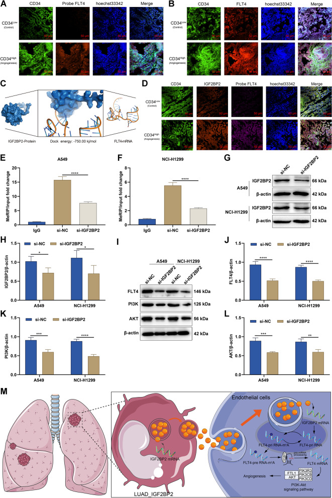
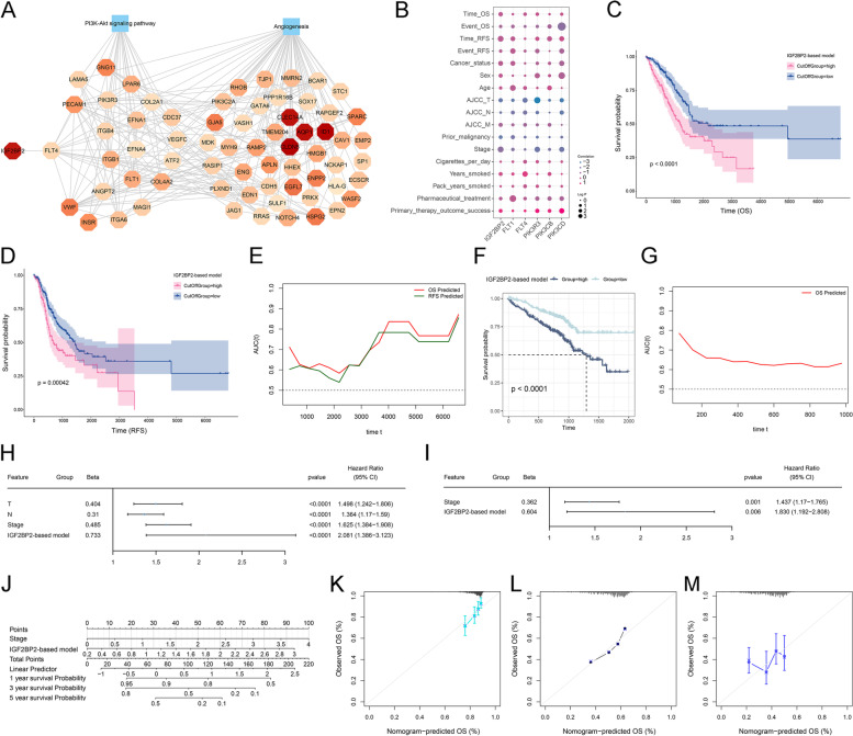
Results: We constructed a global single-cell landscape of 93,610 cells from primary and metastatic LUAD and found that IGF2BP2 was specifically expressed both in a LUAD cell subpopulation (termed as LUAD_IGF2BP2), and an endothelial cell subpopulation (termed as En_IGF2BP2). The LUAD_IGF2BP2 subpopulation progressively formed and dominated the ecology of metastatic LUAD during metastatic evolution. IGF2BP2 was preferentially secreted by exosomes in the LUAD_IGF2BP2 subpopulation, which was absorbed by the En_IGF2BP2 subpopulation in the tumor microenvironment. Subsequently, IGF2BP2 improved the RNA stability of FLT4 through m6A modification, thereby activating the PI3K-Akt signaling pathway, and eventually promoting angiogenesis and metastasis. Analysis of clinical data showed that IGF2BP2 was linked with poor overall survival and relapse-free survival for LUAD patients.

Conclusions: Overall, these findings provide a novel insight into the multicellular ecosystems of primary and metastatic LUAD, and demonstrate that a specific LUAD_IGF2BP2 subpopulation is a key orchestrator promoting angiogenesis and metastasis, with implications for the gene regulatory mechanisms of LUAD metastatic evolution, representing themselves as potential antiangiogenic targets.

Keywords: Angiogenesis; Exosomes; IGF2BP2; Lung adenocarcinoma; Metastasis; N6-methyladenosine; Single-cell RNA sequencing.


Conflict of interest statement

The authors declare no competing interests.

Figures

Fig. 1
Fig. 1
Construction of a global single-cell landscape of primary and metastatic LUAD. A Multiplex immunofluorescence map demonstrating CD34 expression in primary and metastatic LUAD tissues. Bar, 50 μm. B Flow chart of this study. The global single-cell landscape of primary and metastatic LUAD was constructed based on single-cell technology. C The single-cell atlas mapping cell types. D Bubble map showing cell marker genes for cell annotations. E Heat map showing copy number variation results to further validate the accuracy of cell annotation in the presence of multi-copy or low-copy number events in LUAD cells. F The difference in cell abundance between control, primary and metastatic LUAD
Fig. 2
Fig. 2
Clonal evolutionary trajectory of metastatic LUAD cells. A The single cell atlas showing tumor cell subpopulations. B Dotted line graph showing the ratio of distinct tumor cell subpopulations in control, primary and metastatic LUAD. C The single-cell atlas mapping the expression of FAM83A and IGF2BP2 in tumor cells. D Fluorescence assay-fluorescent probe assay showing IGF2BP2 in LUAD tissues with high or low CD34 expression. Bar, 50 μm. E Volcano map showing differentially expressed genes in malignant cells and their subpopulations between metastatic and primary LUAD. F The single-cell atlas mapping the pseudotime values and evolutionary trajectory. G Evolutionary tree showing the evolutionary trajectory of tumor cells. H Heat map-pathway showing signaling pathways involved in temporally relevant genes
Fig. 3
Fig. 3
Endothelial cell landscape in metastatic LUAD. A The single-cell atlas showing endothelial cell subpopulations. B Dotted line graph showing changes in abundance of endothelial cell subpopulations in control, primary and metastatic LUAD. C The single-cell atlas mapping the expression of IGF2BP2 in endothelial cells. D Immunofluorescence assay-fluorescent probe assay showing IGF2BP2 expression in endothelial cell subpopulations with high and low CD34 expression. Bar, 50 μm. E Shared markers of specific cell subpopulations (LUAD_IGF2BP2 and En_IGF2BP2). F Cluster-bubble plots demonstrating biological functions shared by specific cell subpopulations. G Cluster-bubble plots demonstrating KEGG pathways shared by specific cell subpopulations. H GSEA plots demonstrating the significant activation of biological signals related to secretion, secretion and synthesis of vesicles and phagocytosis of contents diffusion in specific cell subpopulations En_IGF2BP2 (left) and LUAD_IGF2BP2 (right)
Fig. 4
Fig. 4
Targeting IGF2BP2 mitigates migration, invasion, and angiogenesis in LUAD cells. A Selection of the optimal siRNAs targeting IGF2BP2 through RT-qPCR. B, C Verification of IGF2BP2 mRNA expression in A549 and NCI-H1299 cells after transfection with si-NC or si-IGF2BP2. D-F Wound healing scratches at 0 h, 24 h, and 48 h for A549 and NCI-H1299 cells transfected with si-NC or si-IGF2BP2. Bar, 200 μm. G, H Transwell assay for detecting invasive A549 and NCI-H1299 cells after transfection with si-NC or si-IGF2BP2. Bar, 50 μm. I-K Images of HUVECs cultured with conditioned medium from A549 cells transfected with si-NC or si-IGF2BP2, and quantification of branch points and capillary length. Bar, 200 μm. *P < 0.05; **P < 0.01; ***P < 0.001; ****P < 0.0001
Fig. 5
Fig. 5
Overexpression of IGF2BP2 in endothelial cells activates the PI3K-Akt signaling to promote angiogenesis. A Circle diagram showing the relationships between LUAD_IGF2BP2 ligand genes and En_IGF2BP2 receptor genes. The gene colors characterize their logFC and the pathway colors are aligned with the circles. The first loop genes are shared markers for the LUAD_IGF2BP2 and the En_IGF2BP2 subpopulations, the second loop is a non-shared marker, and the third and fourth loops are signaling pathways. B Violin diagram showing the expression pattern of exosomal markers in the LUAD subpopulations. C Pathway map showing the PI3K-Akt signaling pathway activated in the En_IGF2BP2 subpopulation
Fig. 6
Fig. 6
IGF2BP2 activates the PI3K-Akt signaling pathway through mediating the m6A modification of FLT4. A Fluorescent probe assay for detecting RNA levels of FLT4 in focal endothelial cells of LUAD patients with high or low CD34 expression. Bar, 50 μm. B Multiplex immunofluorescence assay for measuring protein levels of FLT4 in focal endothelial cells of LUAD patients with high or low CD34 expression. Bar, 50 μm. C Molecular docking for predicting the docking potential of IGF2BP2 protein with mRNA of FLT4. D Multiplex immunofluorescence assay for verifying protein levels of IGF2BP2 and RNA levels of FLT4 in focal endothelial cells of LUAD patients with high or low CD34 expression. Bar, 50 μm. E, F The m.6A levels of FLT4 in A549 and NCI-H1299 cells after transfection with si-NC or si-IGF2BP2. G, H IGF2BP2 protein levels in A549 and NCI-H1299 cells with si-NC or si-IGF2BP2 transfection. I-L FLT4, PI3K, and AKT expression levels in A549 and NCI-H1299 cells with si-NC or si-IGF2BP2 transfection. M The diagram for IGF2BP2-mediated FLT4-PI3K-Akt signaling pathway in regulating angiogenesis during LUAD. *P < 0.05; **P < 0.01; ***P < 0.001; ****P < 0.0001
Fig. 7
Fig. 7
Development of the IGF2BP2-based prognostic scoring system for LUAD. A The global regulatory network of IGF2BP2-FLT4-PI3K-Akt signaling-angiogenesis. B Bubble plots demonstrating the correlation between the IGF2BP2-FLT4-PI3K-Akt signaling-angiogenesis key genes and clinical indicators in the TCGA-LUAD dataset. C, D Survival curves of OS and RFS outcomes for the low and high-score patients stratified by the IGF2BP2-based model in the TCGA-LUAD dataset. E Time-independent ROC curves for evaluating the performance of the IGF2BP2-based model in predicting OS and RFS outcomes in the TCGA-LUAD dataset. F, G External validation of OS analysis and time-independent ROC curves in the GSE72094 dataset. H, I Uni- and multivariate cox regression results on the IGF2BP2-based model and clinical parameters with patient survival in the TCGA-LUAD dataset. J Construction of the nomogram composed of the IGF2BP2-based model and stage. K-M Calibration curves for the nomogram-predicted and actual one-, three- and five-year OS outcomes

Similar articles

Cited by

References

    1. Xia C, Dong X, Li H, Cao M, Sun D, He S, Yang F, Yan X, Zhang S, Li N, Chen W. Cancer statistics in China and United States, 2022: profiles, trends, and determinants. Chin Med J (Engl) 2022;135:584–590. doi: 10.1097/CM9.0000000000002108. - DOI - PMC - PubMed
    1. Gridelli C, Rossi A, Carbone DP, Guarize J, Karachaliou N, Mok T, Petrella F, Spaggiari L, Rosell R. Non-small-cell lung cancer. Nat Rev Dis Primers. 2015;1:15009. doi: 10.1038/nrdp.2015.9. - DOI - PubMed
    1. Kim N, Kim HK, Lee K, Hong Y, Cho JH, Choi JW, Lee JI, Suh YL, Ku BM, Eum HH, et al. Single-cell RNA sequencing demonstrates the molecular and cellular reprogramming of metastatic lung adenocarcinoma. Nat Commun. 2020;11:2285. doi: 10.1038/s41467-020-16164-1. - DOI - PMC - PubMed
    1. Inamura K. Clinicopathological Characteristics and Mutations Driving Development of Early Lung Adenocarcinoma: Tumor Initiation and Progression. Int J Mol Sci. 2018;19(4):1259. doi: 10.3390/ijms19041259. - DOI - PMC - PubMed
    1. Siegel RL, Miller KD, Jemal A. Cancer statistics, 2018. CA Cancer J Clin. 2018;68:7–30. doi: 10.3322/caac.21442. - DOI - PubMed

Publication types

Substances