

Evaluation of diagnostic accuracy of CBCT and intraoral radiography for proximal caries detection in the presence of different dental restoration materials
- PMID: 37353807
- PMCID: PMC10290356
- DOI: 10.1186/s12903-023-02954-8
Evaluation of diagnostic accuracy of CBCT and intraoral radiography for proximal caries detection in the presence of different dental restoration materials
Abstract
Purpose: This study aimed to assess the diagnostic accuracy of cone-beam computed tomography (CBCT) and digital intraoral radiography for the detection of proximal caries adjacent to amalgam, e.max porcelain, and metal-ceramic restorations (MCRs).
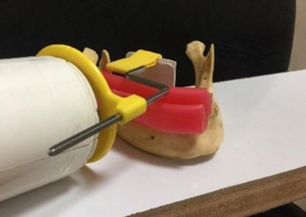
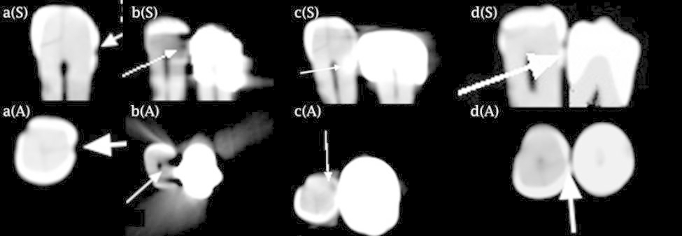
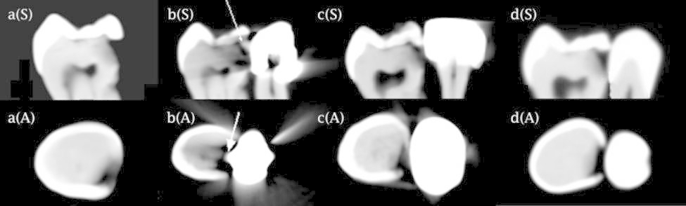
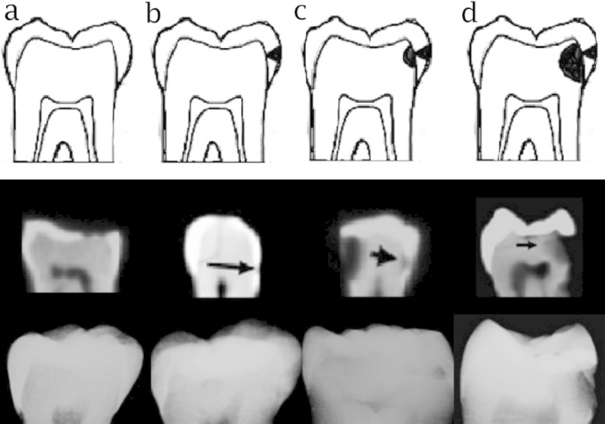
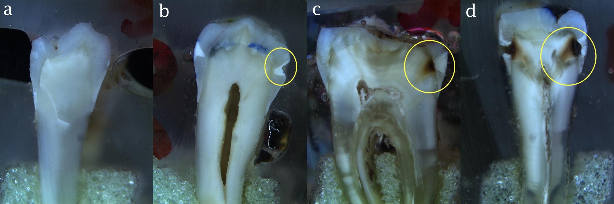
Materials and methods: Parallel intraoral radiographs were obtained from 40 posterior teeth using PSP sensors. To obtain CBCT scans, the teeth were first radiographed alone, and were then positioned next to a tooth with an amalgam restoration, MCR, and e.max porcelain crown, and radiographed again. Two blinded observers scored radiographs using a four-point scale (0: absence of proximal caries, 1: enamel caries, 2: carious lesion extending to the outer half of dentin, 3: carious lesion extending to the inner half of dentin). Tooth sections were made, and the grade of caries was determined under a light microscope at x12 magnification. The sensitivity, specificity, and accuracy of CBCT and intraoral radiographs were then calculated.
Results: Artifact-free CBCT scans and intraoral radiographs had the highest diagnostic accuracy (0.826 and 0.657, respectively) while CBCT images of the teeth next to the amalgam restorations (0.526) had the lowest accuracy. The diagnostic accuracy of CBCT images of the teeth next to the porcelain crowns and MCRs was 0.613 and 0.601, respectively.
Conclusion: Artifact-free CBCT images had higher diagnostic accuracy than intraoral radiography for the detection of all grades of proximal caries. The diagnostic accuracy of CBCT images of teeth adjacent to amalgam, porcelain, and MCRs was lower compared to intraoral radiographs and artifact-free CBCT images.
Keywords: Artifact; Cone-Beam Computed Tomography; Dental Porcelain; Digital radiography.
© 2023. The Author(s).
Conflict of interest statement
The authors declare that they have no competing interests.
Figures
Similar articles
-
Detection accuracy of proximal caries by phosphor plate and cone-beam computerized tomography images scanned with different resolutions.Clin Oral Investig. 2012 Aug;16(4):1015-21. doi: 10.1007/s00784-011-0599-7. Epub 2011 Jul 30. Clin Oral Investig. 2012. PMID: 21805053
-
The impact of different dental restorations on detection of proximal caries by cone beam computed tomography.Clin Oral Investig. 2022 Mar;26(3):2413-2420. doi: 10.1007/s00784-021-04207-w. Epub 2021 Oct 3. Clin Oral Investig. 2022. PMID: 34601634
-
Accuracy of Cone-beam Computed Tomography and Extraoral Bitewings Compared to Intraoral Bitewings in Detection of Interproximal Caries.J Contemp Dent Pract. 2020 Dec 1;21(12):1361-1367. J Contemp Dent Pract. 2020. PMID: 33893259
-
Panoramic radiography in dental diagnostics.Swed Dent J Suppl. 1996;119:1-26. Swed Dent J Suppl. 1996. PMID: 8971997 Review.
-
Performance of the caries diagnosis feature of intraoral scanners and near-infrared imaging technology-A narrative review.J Prosthodont. 2023 Dec;32(S2):114-124. doi: 10.1111/jopr.13770. Epub 2023 Sep 24. J Prosthodont. 2023. PMID: 37701946 Review.
Cited by
-
Sivan classification system for diagnosis of jaw lesions based on visual volumetric analysis of 3-dimensional cone-beam computed tomographic images.Sci Rep. 2024 Dec 30;14(1):32138. doi: 10.1038/s41598-024-83974-4. Sci Rep. 2024. PMID: 39738676 Free PMC article.
-
Dental Caries Detection and Classification in CBCT Images Using Deep Learning.Int Dent J. 2024 Apr;74(2):328-334. doi: 10.1016/j.identj.2023.10.003. Epub 2023 Nov 7. Int Dent J. 2024. PMID: 37940474 Free PMC article.
-
A real-time interactive restoration system for intraoral digital videos using segment anything model.Digit Health. 2024 Aug 5;10:20552076241269536. doi: 10.1177/20552076241269536. eCollection 2024 Jan-Dec. Digit Health. 2024. PMID: 39108255 Free PMC article.
-
Performance of a novel direct-conversion intraoral sensor on the assessment of caries-like lesions - an ex-vivo comparison with conventional (scintillator-dependent) intraoral sensors.Clin Oral Investig. 2025 Jun 3;29(6):329. doi: 10.1007/s00784-025-06400-7. Clin Oral Investig. 2025. PMID: 40456960
-
FDTooth: Intraoral Photographs and CBCT Images for Fenestration and Dehiscence Detection.Sci Data. 2025 Jun 14;12(1):1007. doi: 10.1038/s41597-025-05348-3. Sci Data. 2025. PMID: 40517159 Free PMC article.
References
-
- White SC, Pharoah MJ. Oral Radiology Principles and Interpretation. 6th ed. Mosby; 2009.
Publication types
MeSH terms
Substances
LinkOut - more resources
Full Text Sources
Medical
Miscellaneous
