Review
. 2023 Jun 23;27(1):247.
doi: 10.1186/s13054-023-04532-4.

Respiratory challenges and ventilatory management in different types of acute brain-injured patients

Affiliations
Review

Respiratory challenges and ventilatory management in different types of acute brain-injured patients

S Frisvold et al. Crit Care. .

Abstract

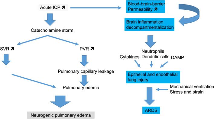
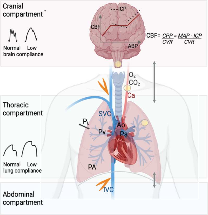
Acute brain injury (ABI) covers various clinical entities that may require invasive mechanical ventilation (MV) in the intensive care unit (ICU). The goal of MV, which is to protect the lung and the brain from further injury, may be difficult to achieve in the most severe forms of lung or brain injury. This narrative review aims to address the respiratory issues and ventilator management, specific to ABI patients in the ICU.

Keywords: Acute brain injury; Acute respiratory distress syndrome; Cerebral autoregulation; Lung protective ventilation; Mechanical ventilation; Neurogenic pulmonary edema.


Conflict of interest statement

The authors declare that they have no competing interests.

Figures

Fig. 1
Fig. 1
Pathways of acute lung injury directly related to acute brain injury. High intracranial pressure (ICP) can promote two different sequences of events that end up into neurogenic pulmonary edema or acute respiratory distress syndrome (ARDS). Both of them may coexist in a given patient. SVR: systemic vascular resistance, PVR: pulmonary vascular resistance, LV: left ventricle, DAMP: damage-associated molecules pattern
Fig. 2
Fig. 2
Schematic representation of the lung–brain interactions. During positive pressure mechanical ventilation, cerebral blood flow (CBF) can be reduced from different sources. The transmission of airway pressure to the cardiovascular structures depends on the pleural pressure and thus on the transpulmonary pressure (PL) and lung compliance. With normal lung compliance, the higher the airway pressure, the higher the right atrial pressure, which can lead to a reduction in venous return (orange flash). Increase in abdominal pressure counteracts this effect in normal conditions. Increased tidal volume increases pulmonary venous pressure (Pv). These changes result in lower right ventricular ejection volume, and thus, cardiac output (CO) will decrease. CO reduction is limited by the fact that the increased intrathoracic pressure will decrease the left ventricle afterload. Despite changes in CO and arterial pressure, cerebral autoregulation maintains CBF and intracranial pressure (ICP) within a certain range of arterial pressure. However, ICP is highly dependent on venous outflow from the cranial cavity. Positive pressure ventilation with increased right atrial pressure can reduce venous outflow from the cranial cavity and thereby increase ICP. In patients with impaired pulmonary compliance (i.e., severe acute respiratory distress syndrome), the effects of positive pressure mechanical ventilation on alveolar pressure (PA) and PL are often attenuated. Hypoxemia (low PaO2) and hypercapnia (high PaCO2) both increase pulmonary artery pressure (Pa) and pulmonary vascular resistance, thereby increasing right ventricular afterload. Alterations in PaCO2, PaO2 and hydrogen ion also trigger chemoreceptors (yellow circles) to send signals to the respiratory center to regulate respiratory drive. At the level of cerebral circulation, hypercapnia increases CBF and hypocapnia has the opposite effect. The interaction between low brain compliance, cerebral autoregulation and different levels of CO2 has not been studied. Ao aorta, PaCO2 partial pressure of carbon dioxide, PaO2 partial pressure of oxygen, Ca carotid artery, cardiac output (CO), CBF cerebral blood flow, CPP cerebral perfusion pressure, CVR cerebral vascular resistance, ICP intracranial pressure, IVC inferior vena cava, MAP mean arterial pressure, Pa pulmonary artery pressure, PA alveolar pressure, Pv pulmonary venous pressure, PaO2 partial pressure of oxygen, PL transpulmonary pressure, SVC superior vena cava

References

    1. Pelosi P, Ferguson ND, Frutos-Vivar F, Anzueto A, Putensen C, Raymondos K, et al. Management and outcome of mechanically ventilated neurologic patients. Crit Care Med. 2011;39(6):1482–1492. doi: 10.1097/CCM.0b013e31821209a8. - DOI - PubMed
    1. Ziaka M, Exadaktylos A. Brain–lung interactions and mechanical ventilation in patients with isolated brain injury. Crit Care. 2021;25(1):1–10. doi: 10.1186/s13054-021-03778-0. - DOI - PMC - PubMed
    1. Robba C, Poole D, McNett M, Asehnoune K, Bösel J, Bruder N, et al. Mechanical ventilation in patients with acute brain injury: recommendations of the European Society of Intensive Care Medicine consensus. Intensive Care Med. 2020;46(12):2397–2410. doi: 10.1007/s00134-020-06283-0. - DOI - PMC - PubMed
    1. Meng L, Gelb AW. Regulation of cerebral autoregulation by carbon dioxide. Anesthesiology. 2015;122:196–205. doi: 10.1097/ALN.0000000000000506. - DOI - PubMed
    1. North JB, Jennett S. Abnormal breathing patterns associated with acute brain damage. Arch Neurol. 1974;31(5):338–344. doi: 10.1001/archneur.1974.00490410086010. - DOI - PubMed