

Investigation of ocular involvement in patients with Fabry disease
- PMID: 37354009
- PMCID: PMC10291927
- DOI: 10.1080/07853890.2023.2226909
Investigation of ocular involvement in patients with Fabry disease
Abstract
Purpose: To investigate ocular abnormalities in Fabry disease (FD).
Methods: Forty-five patients with FD diagnosed by genetic analysis were enrolled in a single medical centre. The following ocular examinations were performed: slit-lamp examination, ophthalmic fundus imaging, in vivo confocal microscopy (IVCM) and optical coherence tomography (OCT). The prevalences of typical abnormalities in the cornea, conjunctiva and retina were recorded; their differences between hemizygote and heterozygote were compared.
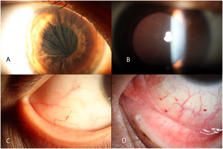
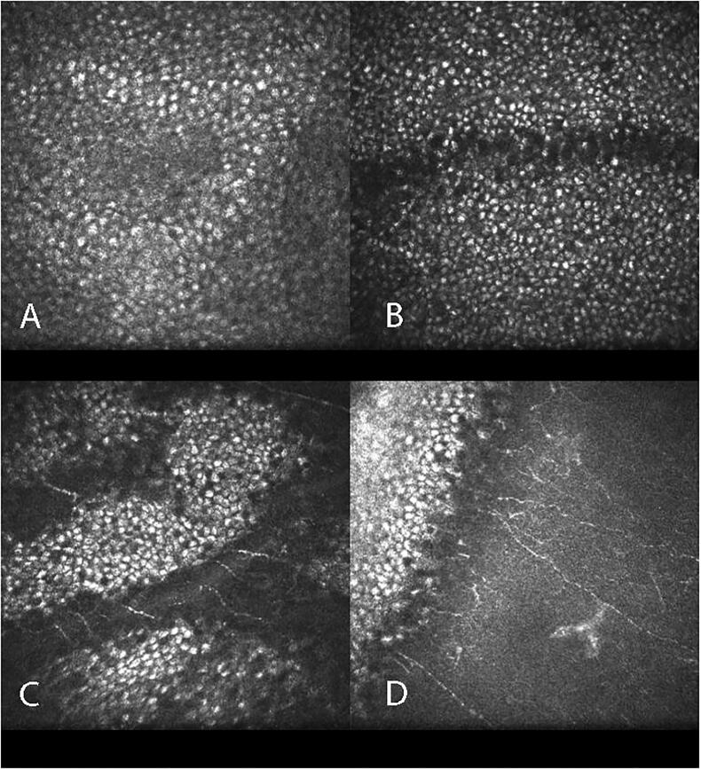
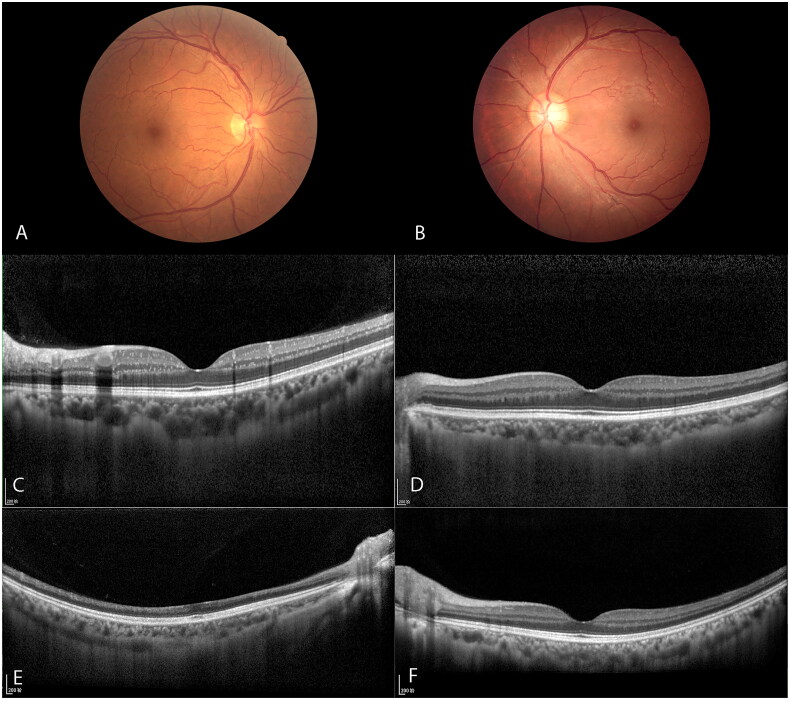
Results: In this study, the prevalence of corneal verticillata was 97.8% (44/45). Corneal examination with IVCM demonstrated hyper-reflective intracellular inclusions located within basal epithelial cells. Conjunctival vessel malformations were observed in 64.4% (29/45) of patients, and retinal vessel tortuosity was observed in 62.2% (28/45) of patients. OCT revealed many strong hyper-reflective foci (HRF) in the inner retinal layer (in 66.7% [30/45] of patients); these foci may represent retinal vascular plexi. The prevalences of conjunctival vessel malformation, retinal vessel tortuosity and HRF were higher in hemizygote than in heterozygote.
Conclusions: Corneal verticillata, HRF on OCT, conjunctival vessel malformation and retinal vessel tortuosity exhibit high prevalence in patients with FD. These ocular manifestations are characteristic and easily accessible; thus, they should be considered diagnostic criteria for FD.
Keywords: Fabry disease; corneal verticillata; optical coherence tomography; retinal vessel tortuosity.
Plain language summary
The main ocular features of the patients with FD are corneal verticillata, HRF on OCT, conjunctival vessel malformation and retinal vessel tortuosity.These ocular manifestations should be considered as the diagnostic criteria of FD.
Conflict of interest statement
No potential conflict of interest was reported by the author(s).
Figures
Similar articles
-
Ocular Manifestations of Fabry Disease: Report from a Tertiary Eye Care Center in Türkiye.Turk J Ophthalmol. 2024 Jun 28;54(3):127-132. doi: 10.4274/tjo.galenos.2024.09482. Turk J Ophthalmol. 2024. PMID: 38940325 Free PMC article.
-
Cornea verticillata in Fabry disease: a comparative study between slit-lamp examination and in vivo corneal confocal microscopy.Br J Ophthalmol. 2020 May;104(5):718-722. doi: 10.1136/bjophthalmol-2019-314249. Epub 2019 Aug 10. Br J Ophthalmol. 2020. PMID: 31401555
-
Retinal hyperreflective foci in Fabry disease.Orphanet J Rare Dis. 2019 Dec 26;14(1):296. doi: 10.1186/s13023-019-1267-2. Orphanet J Rare Dis. 2019. PMID: 31878969 Free PMC article.
-
Ophthalmic Manifestations in Fabry Disease: Updated Review.J Pers Med. 2023 May 27;13(6):904. doi: 10.3390/jpm13060904. J Pers Med. 2023. PMID: 37373893 Free PMC article. Review.
-
Ophthalmological manifestations of Fabry disease.In: Mehta A, Beck M, Sunder-Plassmann G, editors. Fabry Disease: Perspectives from 5 Years of FOS. Oxford: Oxford PharmaGenesis; 2006. Chapter 26. In: Mehta A, Beck M, Sunder-Plassmann G, editors. Fabry Disease: Perspectives from 5 Years of FOS. Oxford: Oxford PharmaGenesis; 2006. Chapter 26. PMID: 21290696 Free Books & Documents. Review.
Cited by
-
The vestibular and oculomotor dysfunction in Fabry disease: a cohort study in China.Ann Med. 2025 Dec;57(1):2453626. doi: 10.1080/07853890.2025.2453626. Epub 2025 Jan 25. Ann Med. 2025. PMID: 39862133 Free PMC article.
-
CDK4/6 inhibitor-associated vortex keratopathy: a case report.BMC Ophthalmol. 2025 Apr 14;25(1):206. doi: 10.1186/s12886-025-04043-6. BMC Ophthalmol. 2025. PMID: 40229836 Free PMC article.
-
Deep learning assisted retinal microvasculature assessment and cerebral small vessel disease in Fabry disease.Orphanet J Rare Dis. 2025 Apr 3;20(1):158. doi: 10.1186/s13023-025-03627-1. Orphanet J Rare Dis. 2025. PMID: 40181436 Free PMC article.
-
Ocular and confocal manifestations of Mainland Chinese with Fabry disease: a cross-sectional controlled study.Orphanet J Rare Dis. 2025 Aug 10;20(1):417. doi: 10.1186/s13023-025-03940-9. Orphanet J Rare Dis. 2025. PMID: 40784937 Free PMC article.
-
Optical Coherence Tomography (OCT): A Brief Look at the Uses and Technological Evolution of Ophthalmology.Medicina (Kaunas). 2023 Dec 3;59(12):2114. doi: 10.3390/medicina59122114. Medicina (Kaunas). 2023. PMID: 38138217 Free PMC article. Review.
References
-
- Chan B, Adam DN.. A review of Fabry disease. Skin Therapy Lett. 2018;23:1–7. - PubMed