. 2023 Aug;32(8):e4711.
doi: 10.1002/pro.4711.

Investigating the inhibitory property of DM hCT on hCT fibrillization via its relevant peptide fragments

Affiliations

Investigating the inhibitory property of DM hCT on hCT fibrillization via its relevant peptide fragments

Ya-Ping Chuang et al. Protein Sci. 2023 Aug.

Abstract

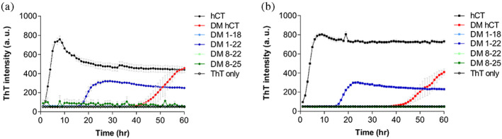
The irreversible aggregation of proteins or peptides greatly limits their bioavailability; therefore, effective inhibition using small molecules or biocompatible materials is very difficult. Human calcitonin (hCT), a hormone polypeptide with 32 residues, is secreted by the C-cells of the thyroid gland. The biological function of this hormone is to regulate calcium and phosphate concentrations in the blood via several different pathways. One of these is to inhibit the activity of osteoclasts; thus, calcitonin could be used to treat osteoporosis and Paget's disease of the bone. However, hCT is prone to aggregation in aqueous solution and forms amyloid fibrils. Salmon and eel calcitonin are currently used as clinical substitutes for hCT. In a previous study, we found that the replacement of two residues at positions 12 and 17 of hCT with amino acids that appear in the salmon sequence can greatly suppress peptide aggregation. The double mutations of hCT (DM hCT) also act as good inhibitors by disrupting wild-type hCT fibrillization, although the inhibition mechanism is not clear. More importantly, we demonstrated that DM hCT is biologically active in interacting with the calcitonin receptor. To further understand the inhibitory effect of DM hCT on hCT fibrillization, we created four relevant peptide fragments based on the DM hCT sequence. Our examination revealed that the formation of a helix of DM hCT was possibly a key component contributing to its inhibitory effect. This finding could help in the development of peptide-based inhibitors and in understanding the aggregation mechanism of hCT.

Keywords: amyloid formation; human calcitonin; mutations; osteoporosis; peptide-based inhibitors.


Conflict of interest statement

The authors declare no conflict of interests.

Figures

FIGURE 1
FIGURE 1
Primary sequence of hCT, DM hCT, and DM hCT‐relevant peptide fragments. hCT, DM hCT, DM 1–18, and DM 1–22 contain a disulfide bridge between Cys 1 and Cys 7. DM hCT and its relevant peptide fragments have two mutation sites at residue 12 and 17 labeled in red. The C‐terminus of all peptides used in this study is amidated.
FIGURE 2
FIGURE 2
ThT monitored amyloid formation kinetics of hCT (black), DM hCT (red), DM 1–18 (light blue), DM 1–22 (blue), DM 8–22 (light green), and DM 8–25 (green) conducted at (a) 20 μM and (b) 30 μM, respectively, in 10 mM phosphate buffer at pH 7.4. Each condition was performed in triplicates.
FIGURE 3
FIGURE 3
ThT monitored amyloid formation kinetics of hCT with DM hCT (red), with DM 1–18 (light blue), with DM 1–22 (blue), with DM 8–22 (light green), and with DM 8–25 (green). The concentration of hCT was fixed at 20 μM. DM hCT and its peptide fragments were added at (a) 20 μM and (b) 30 μM. (c, d) Analysis of the times needed to reach reaction plateau for each experimental condition. N/A represents no ThT intensity monitored within 50 h.
FIGURE 4
FIGURE 4
The CD spectra of (a) DM hCT, (b) DM 1–18, (c) DM 1–22, (d) DM 8–22, and (e) DM 8–25 prepared in 50 mM phosphate buffer at pH 7.4 with the presence of 0% to 40% TFE (form light color to dark color).
FIGURE 5
FIGURE 5
Percent composition of helix estimated by CD fitting software, BeStStel for each condition shown in Figure 4.
FIGURE 6
FIGURE 6
The CD spectra of (a) DM hCT, (b) DM 1–18, (c) DM 1–22, (d) DM 8–22, and (e) DM 8–25 prepared in 50 mM phosphate buffer at pH 7.4 without (black) and with 400 μM DMPG (red).
FIGURE 7
FIGURE 7
(a) ThT monitored amyloid formation kinetics of hCT (black), hCT with 20 μM DM 8–22 (light green) and hCT with 30 μM DM 8–22 (green). (b, c) TEM images of hCT with 20 and 30 μM DM 8–22 at the end of kinetic experiments. Scare bar represents 200 nm. (d) ThT monitored amyloid formation kinetics of hCT (black), hCT with 20 μM DM 8–25 (light green) and hCT with 30 μM DM 8–25 (green). (e, f) TEM images of hCT with 20 and 30 μM DM 8–25 at the end of kinetic experiments. Scare bar represents 200 nm. hCT was prepared at 20 μM in each kinetic assay (10 mM phosphate buffer at pH 7.4 with 30% TFE).
FIGURE 8
FIGURE 8
A schematic diagram of how DM hCT inhibits hCT aggregation. Formation of a stable helix for DM hCT would be an important step which allows it to interact with helical structure of hCT.

Similar articles

Cited by

References

    1. Abedini A, Raleigh DP. A critical assessment of the role of helical intermediates in amyloid formation by natively unfolded proteins and polypeptides. Protein Eng Des Select. 2009a;22(8):453–9. - PMC - PubMed
    1. Abedini A, Raleigh DP. A role for helical intermediates in amyloid formation by natively unfolded polypeptides? Phys Biol. 2009b;6(1):15005. - PMC - PubMed
    1. Amodeo P, Motta A, Strazzullo G, Castiglione Morelli MA. Conformational flexibility in calcitonin: the dynamic properties of human and salmon calcitonin in solution. J Biomol NMR. 1999;13(2):161–74. - PubMed
    1. Andreotti G, Méndez BL, Amodeo P, Castiglione Morelli MA, Nakamuta H, Motta A. Structural determinants of salmon calcitonin bioactivity: the role of the leu‐based amphipathic α‐helix. J Biol Chem. 2006;281(34):24193–203. - PubMed
    1. Andreotti G, Vitale RM, Avidan‐Shpalter C, Amodeo P, Gazit E, Motta A. Converting the highly amyloidogenic human calcitonin into a powerful fibril inhibitor by three‐dimensional structure homology with a non‐amyloidogenic analogue. J Biol Chem. 2011;286(4):2707–18. - PMC - PubMed

Publication types

LinkOut - more resources