. 2023 Jul 24;33(14):3002-3010.e6.
doi: 10.1016/j.cub.2023.05.071. Epub 2023 Jun 23.

Systemic coagulopathy promotes host lethality in a new Drosophila tumor model

Affiliations

Systemic coagulopathy promotes host lethality in a new Drosophila tumor model

Tsai-Ching Hsi et al. Curr Biol. .

Abstract

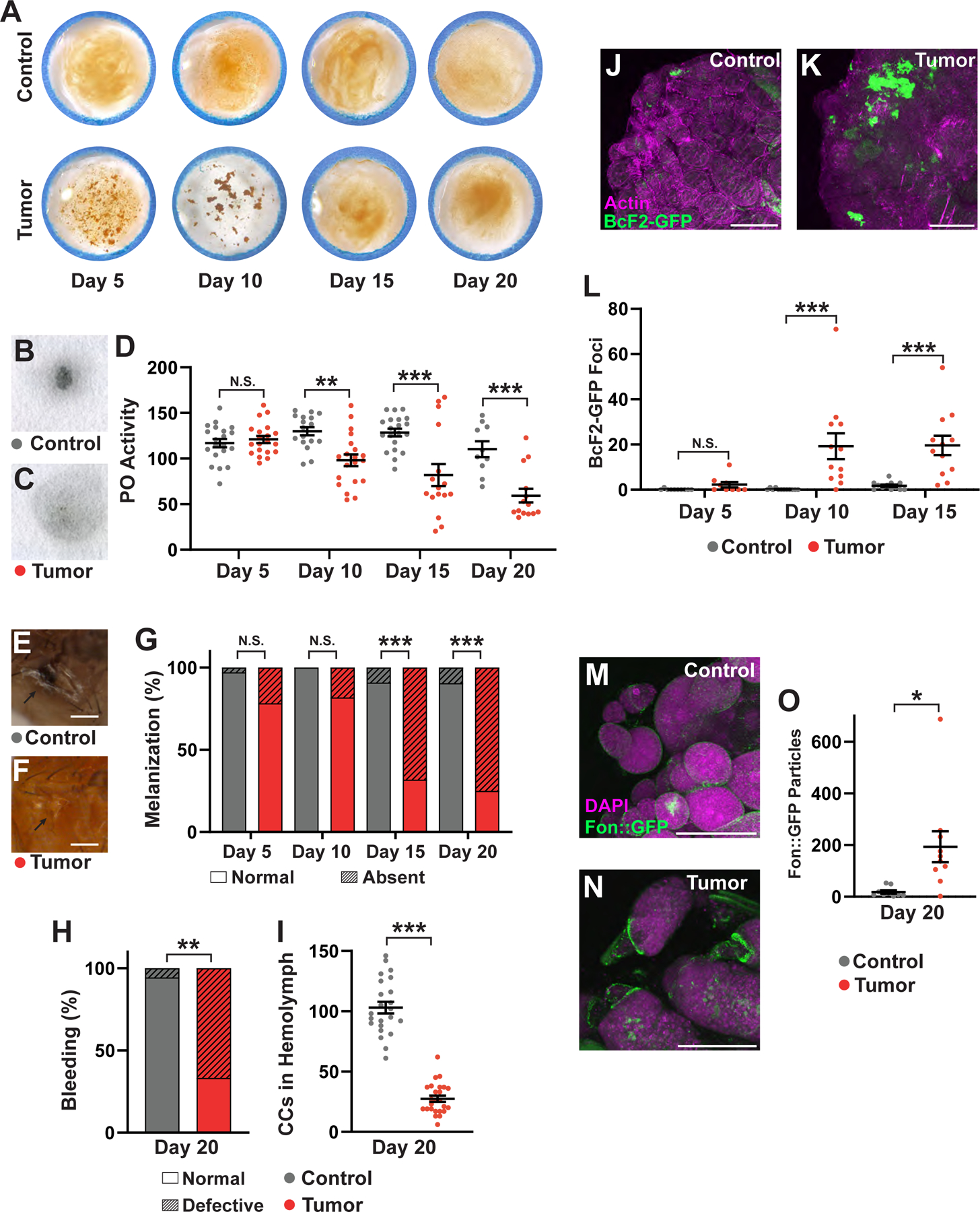
Malignant tumors trigger a complex network of inflammatory and wound repair responses, prompting Dvorak's characterization of tumors as "wounds that never heal."1 Some of these responses lead to profound defects in blood clotting, such as disseminated intravascular coagulopathy (DIC), which correlate with poor prognoses.2,3,4 Here, we demonstrate that a new tumor model in Drosophila provokes phenotypes that resemble coagulopathies observed in patients. Fly ovarian tumors overproduce multiple secreted components of the clotting cascade and trigger hypercoagulation of fly blood (hemolymph). Hypercoagulation occurs shortly after tumor induction and is transient; it is followed by a hypocoagulative state that is defective in wound healing. Cellular clotting regulators accumulate on the tumor over time and are depleted from the body, suggesting that hypocoagulation is caused by exhaustion of host clotting components. We show that rescuing coagulopathy by depleting a tumor-produced clotting factor improves survival of tumor-bearing flies, despite the fact that flies have an open (non-vascular) circulatory system. As clinical studies suggest that lethality in patients with high serum levels of clotting components can be independent of thrombotic events,5,6 our work establishes a platform for identifying alternative mechanisms by which tumor-driven coagulopathy triggers early mortality. Moreover, it opens up exploration of other conserved mechanisms of host responses to chronic wounds.

Keywords: Tumor-host; carcinoma; clotting; coagulopathy; drosophila; innate immunity; ovarian cancer; paraneoplasia; pre-clinical disease model; wound response.


Conflict of interest statement

Declaration of interests The authors declare no competing interests.

Figures

Figure 1.
Figure 1.. A novel ovarian carcinoma model to study paraneoplasias in Drosophila.
(A) Schematic of Drosophila ovarian carcinoma (OC) induction in adult flies. (B, C) OC tumors exhibit increased anti-phospho-histone H3 staining (green, Max Z-projections), quantitated in (D, n-values: Control=19, Tumor=17). (E, F) Apical SAS::Venus (yellow) and lateral anti-Dlg staining (cyan) reveal disruption of cellular organization in transformed follicle epithelial cells (n-values: Control=3, Tumor=7). (G, H) OC tumors fail to differentiate into the mature Hnt-positive (red), FasIII-negative (cyan) follicles of control; numbers in G represent follicle stages (n-values: Control=10, Tumor=14). (I) Flies carrying OCs (n=176) have greatly reduced survival compared to control, non-tumor-bearing flies (n=338). Tumor-bearing flies exhibit substantial fluid accumulation as indicated by abdomen distention (J, K, n-values: Control=39, Tumor=70) and quantification of water weight (L, n-values: Control=9, Tumor=12). (M, N) Lipid staining (red) shows decreased fat tissue in OC flies (Control mean grey value=767.13±28.01 au, n=10; Tumor mean grey value=458.97±69.50 au, n=18; p=0.0011). (O) OC flies exhibit increased permeability of the blood-brain barrier (n-values: Control=19, Tumor=18). Scale Bars = 100μm (B, C), 50μm (G, H), 25μm (E, F), 1mm (J, K), 250μm (M, N); Error bars = S.E.M; Two-tailed t-test (D, L, O) and log-rank test (I) used to determine significance; *p < .05, ***p < .0005, ****p < .00005. Dots in (L) represent pools of 4–5 flies, (O) represent individual flies. See also Figure S1, S2 and Table S1, S2.
Figure 2.
Figure 2.. OCs differentially express clotting factors and regulators, among a number of other secreted proteins.
(A) OCs exhibit heightened transcription of JNK pathway targets MMP1 (60.97X, p<0.0005) and puc (3.53X, p=3.68×10⁻¹⁵), as well as STAT pathway targets mfas (8.28X, p=6.59×10⁻³²) and SOCS36E (1.52X, p=4.53×10⁻⁴). (B) Increased proportion of OC tumor cells in early differentiation is indicated by enhanced transcript levels of GstS1 (3.45X, 4.73×10⁻¹⁰) and Zfh1 (5.35X, p=5.35×10−38), and the failure to upregulate hnt (−59.71X, p=5.28×10−237). (C) 246 of 3567 transcripts upregulated in OC tumors vs control follicles are predicted to encode secreted proteins. (D) Like other Drosophila tumors, OCs upregulate upd2 (32.58X, p=1.95×10⁻¹⁶) and upd3 (2.68X, p=3.34×10⁻⁶), Pvf2 (173.00X, p=8.82×10−52), Pvf3 (6.41X, p=2.47×10⁻⁵), and ImpL2 (9.54X, p=1.16×10⁻²¹). (E) Expression of many factors associated with hemolymph clotting are increased in OCs compared to wild-type follicle cells: fon (28.58X, p=3.75×10⁻⁵), hml (12.89X, 2.97×10⁻¹⁹), Tg (23.92X, p=1.15×10−69), PPO1 (18.84X, p=0.0296), PPO2 (24.22X, p=0.169), and FBP1 (8.35X, p=0.07). Two biological replicates were generated for RNA-sequencing analysis. Error bars = S.E.M. See also Figure S2 and Table S1, S2.
Figure 3:
Figure 3:. Drosophila ovarian tumors induce multiple defects in clotting.
(A) Bead aggregation assay shows that soft clotting activity, which is low in control adult hemolymph (top), is dramatically increased in OC adult hemolymph (bottom) through day 10 AT. Images are representative of ≥3 biological replicates and do not come from a single time course. PO activity in control (B) and OC flies (C) measured by L-DOPA blot reactivity. (D) Quantification of PO activity reveals decreases in OC flies following 10 days ATI (n≥10 for each group). Thoracic wound response in control (E) and OC flies (F). (G) Percentage of flies able to melanize wounds reveals strong differences on days 15 and 20 ATI (n≥22 for each group). (H) Hypocoagulation of OC flies, demonstrated by ability to extract hemolymph from thoracic wounds after 2 hours (n-values: Control=18, Tumor=9). (I) Compared to control (n=23), BcF2-positive crystal cell counts in hemolymph decrease in tumor-bearing flies (n=25) at day 20 ATI. Only a few BcF2-positive cells are found on control ovaries (J) compared to several dozen on ovarian tumors day 15 ATI (K); quantitated in (L, n≥10 for each group). A Fon::GFP fusion protein expressed in control ovaries (M) shows diffuse signal, whereas the same fusion protein expressed in OC cells shows particulate aggregation (N). Quantitated in (O, n ≥9). (J, K, M, N) show maximum-projected images. Scale Bars = 250μm (E,F), 100μm (I,J,M,N); Error bars = S.E.M.; Two-way ANOVA (D, L), Fisher’s Exact test (G, H) and student’s T-test (I, O) used to determine significance; *p < .05, **p < .005, ***p < .0005, N.S. = not significant. Dots in (D) and (I) represent individual flies, (L) and (O) represent individual ovaries. See also Figure S3, Table S2.
Figure 4:
Figure 4:. Tumor-secreted Fondue drives coagulopathies and early mortality.
(A) Early mortality induced by OC tumors is significantly reduced upon Fon depletion (n-values: Ctrl RNAi=160, fon RNAi=142). Compared to control RNAi depletion in OC cells (B), Fon depletion in OC cells (C) rescues the strong hypercoagulation displayed by tumor-bearing flies, revealed by the bead aggregation assay and quantitated in (D, n-values: Ctrl RNAi=6, fon RNAi=8). Compared to control RNAi expression in OC cells (E), Fon depletion in OC cells (F) also ameliorates the loss of PO activity, quantitated in (G, n-values: Ctrl RNAi=18, fon RNAi=40), defects in bleeding (H, n-values: Ctrl RNAi=8, fon RNAi=45) and wound melanization (I-K, n-values: Ctrl RNAi=75, fon RNAi=122). Scale Bars = 25μm; Error bars = S.E.M.; Log-rank test (A), student’s T-test (D, G), and Fisher’s Exact test (H, K) used to determine significance; *p < .05, ***p < .0005. Dots in (D) represent 2 ul of hemolymph pooled from ≥20 flies, (G) represent individual flies. See also Figure S1, S3, S4 and Table S1, S2.

References

    1. Dvorak HF (1986). Tumors: Wounds that do not heal. N Engl J Med 315, 1650–1659. - PubMed
    1. Levi M, and ten Cate H (1999). Disseminated Intravascular Coagulation. N Engl J Med 341, 586–592. - PubMed
    1. Levi M (2009). Disseminated intravascular coagulation in cancer patients. Best Pract Res Clin Haematol 22, 129–136. 10.1016/j.beha.2008.12.005. - DOI - PubMed
    1. Khorana AA (2010). Venous thromboembolism and prognosis in cancer. Thromb Res 125, 490–493. 10.1016/j.thromres.2009.12.023. - DOI - PMC - PubMed
    1. Krenn-Pilko S, Langsenlehner U, Stojakovic T, Pichler M, Gerger A, Kapp KS, and Langsenlehner T (2015). An elevated preoperative plasma fibrinogen level is associated with poor disease-specific and overall survival in breast cancer patients. Breast 24, 667–672. 10.1016/j.breast.2015.08.003. - DOI - PubMed

Publication types