. 2023;16(4):573-605.
doi: 10.1016/j.jcmgh.2023.06.010. Epub 2023 Jun 22.

Types of Neurons in the Human Colonic Myenteric Plexus Identified by Multilayer Immunohistochemical Coding

Affiliations

Types of Neurons in the Human Colonic Myenteric Plexus Identified by Multilayer Immunohistochemical Coding

Bao Nan Chen et al. Cell Mol Gastroenterol Hepatol. 2023.

Abstract

Background and aims: Gut functions including motility, secretion, and blood flow are largely controlled by the enteric nervous system. Characterizing the different classes of enteric neurons in the human gut is an important step to understand how its circuitry is organized and how it is affected by disease.

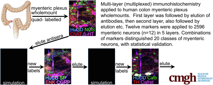
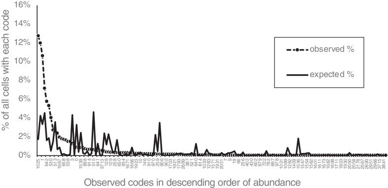
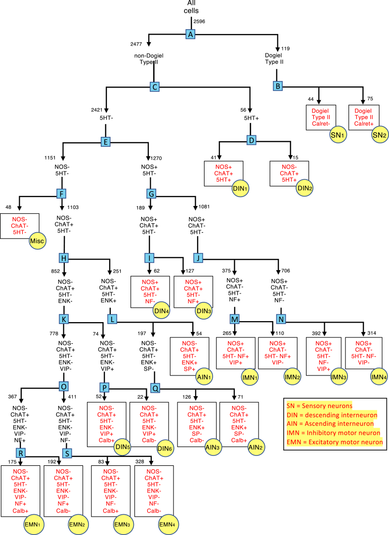
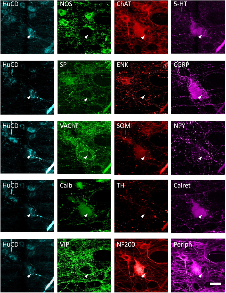
Methods: Using multiplexed immunohistochemistry, 12 discriminating antisera were applied to distinguish different classes of myenteric neurons in the human colon (2596 neurons, 12 patients) according to their chemical coding. All antisera were applied to every neuron, in multiple layers, separated by elutions.

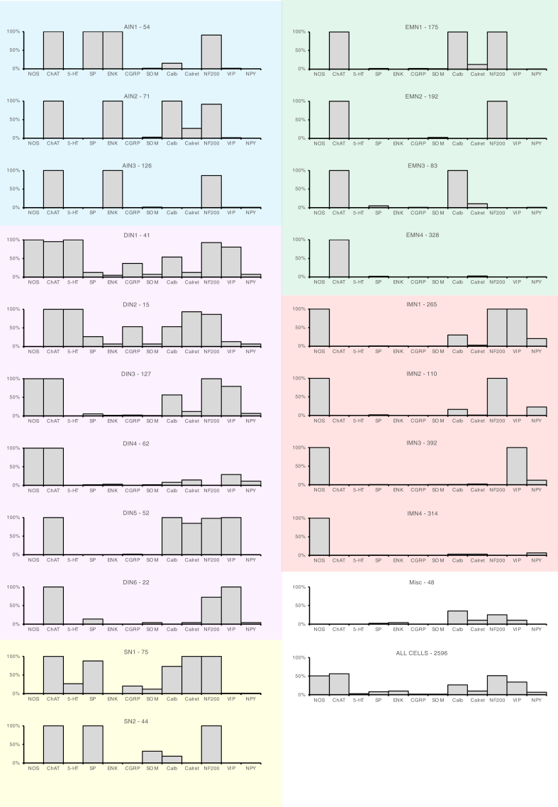
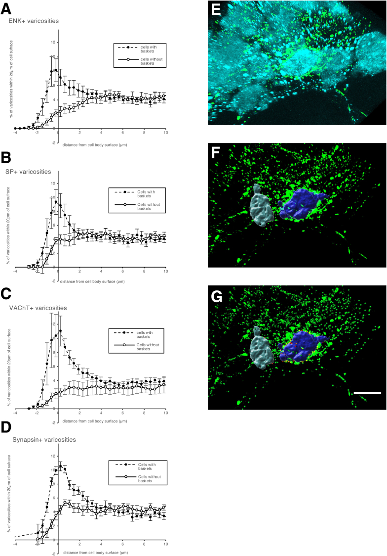
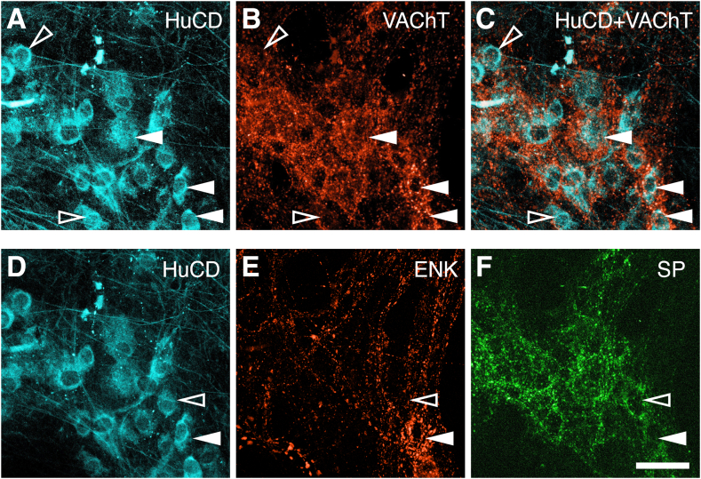
Results: A total of 164 combinations of immunohistochemical markers were present among the 2596 neurons, which could be divided into 20 classes, with statistical validation. Putative functions were ascribed for 4 classes of putative excitatory motor neurons (EMN1-4), 4 inhibitory motor neurons (IMN1-4), 3 ascending interneurons (AIN1-3), 6 descending interneurons (DIN1-6), 2 classes of multiaxonal sensory neurons (SN1-2), and a small, miscellaneous group (1.8% of total). Soma-dendritic morphology was analyzed, revealing 5 common shapes distributed differentially between the 20 classes. Distinctive baskets of axonal varicosities surrounded 45% of myenteric nerve cell bodies and were associated with close appositions, suggesting possible connectivity. Baskets of cholinergic terminals and several other types of baskets selectively targeted ascending interneurons and excitatory motor neurons but were significantly sparser around inhibitory motor neurons.

Conclusions: Using a simple immunohistochemical method, human myenteric neurons were shown to comprise multiple classes based on chemical coding and morphology and dense clusters of axonal varicosities were selectively associated with some classes.

Keywords: Antisera; Classes; Enteric Nervous System; Immunofluorescence.


Figures

None
Graphical abstract
Figure 1
Figure 1
Effects of antibody elution on immunohistochemical labeling. The top row shows typical multiple labeling with HuCD visualized with a biotinylated secondary and AMCA-labeled tertiary. Primaries for the other 3 markers, NOS, ChAT, and 5-HT, were visualized with fluorophore-labeled secondary antisera. The middle row shows the same ganglion after 60 minutes of elution. Note that the HuCD (with biotin-streptavidin complexes) is unaffected but labeling in the other 3 channels is abolished apart from faint autofluorescence of the ganglion and red blood cells. In the third layer, primary antisera to SP, ENK, and CGRP were applied and visualized with fluorophore-coupled secondary antisera, revealing quite different patterns of immunoreactivity from the top row (although ENK and ChAT largely coexist and the 5-HT cell is also immunoreactive for CGRP). Images were taken at exposures shown with false colors applied but no alterations to brightness or contrast.
Figure 2
Figure 2
Markers of myenteric neurons are far from randomly assorted. Each combination of markers was given a numerical code from 0 to 4095 (see Materials and Methods and Results and Table 5). Percentages of cells expressing codes were ordered by descending frequency (dashed line) and compared with expected frequencies calculated if markers randomly combined in their original proportions (solid line). Note that a few classes were far more abundant than expected; these included 2 classes of IMNs (codes 1025 and 1) (n = 12 patients).
Figure 3
Figure 3
Dendrogram summarizing the divisive hierarchical analysis of 2596 cells that distinguished 20 types of neurons in the myenteric plexus of human colon (n = 12). A series of binary divisions was undertaken, based on statistical identification of the most discriminating marker for each population (see Materials and Methods). Red font and yellow circles denote endpoints (classes). Division points are letters in blue boxes. Numbers next to arrows indicate the number of cells at each branch point. The statistical validation of each branch point (blue boxes A–S) is summarized in Table 6.
Figure 4
Figure 4
Identification of Dogiel type II neurons and their coding. A large multiaxonal Dogiel type II cell is clearly identified by NF200 immunoreactivity (fifth row, white arrowhead indicates nucleus). The same cell was immunoreactive for HuCD, ChAT, 5-HT (top row), CGRP (second row), SOM (3rd row), Calret, Calb (fourth row), and peripherin (fifth row) but not for NOS, SP, ENK, NPY, TH, or VIP. The code for this cell was calculated (ChAT = 2, 5-HT = 4, CGRP = 32, SOM = 64, Calb = 128, Calret = 256, NF200 = 512, with all other nonimmunoreactive markers set to 0; this summed to give this cell a code of 998). Micrographs were manually realigned between layers of immunohistochemistry. Similar data were obtained in the 12 patients studied with the full multilayer protocol). Calibration bar (bottom right) = 50 μm.
Figure 5
Figure 5
The 20 types of neurons distinguished in the study differed in the combinations of markers that they expressed. Each graph shows one type of neuron. The x-axis shows the 12 cell body markers and the y-axis shows the proportion of neurons immunoreactive for each marker. In general, 100% or 0% indicates that a marker had been used to discriminate subtypes further up the dendrogram. There were 3 types of AINs (AIN1–3), 6 types of DINs (DIN1–6), 2 intrinsic SNs (multiaxonal Dogiel type II neurons: SN1, SN2), 4 types of EMNs (EMN1–4), 4 IMNs (IMN1–4), and a small population (1.8%) that lacked both NOS and ChAT immunoreactivity (Misc). n = 2596 cells from 12 patients.
Figure 6
Figure 6
Distribution of varicosities relative to cell body surface compared between cells with baskets and with no baskets. (A–D) Distribution of varicosities immunoreactive for ENK, SP, VAChT, and synapsin 1a/1b as 3-dimensional (3D) distance from nerve cell body, for baskets (dashed line) and nonbasket (solid line). Note the large peak of varicosities within 2 μm of the cell surface in baskets that was absent in cells that did not receive baskets (y-axis data are normalized). (E–G) 3D rendering of myenteric nerve cell bodies (HuCD+) and ENK+ varicosities (green), with 2 cells rendered in 3D (F) with closely apposing varicosities (within 2 μm) shown by red spheres (G); these are numerous around the purple soma (surrounded by a basket of varicosities) but less so around the gray cell (no basket). This shows that visually identified baskets make a higher proportion of close appositions to myenteric nerve cell bodies than is the case for cells not surrounded by identifiable baskets. (A–D) Data from 24 cells from 2 patients. (E–G) Calibration bar = 20 μm.
Figure 7
Figure 7
Baskets of labeled axonal varicosities surround some but not all myenteric nerve cell bodies. (A, D) Left micrographs show HuCD somata (cyan) that persisted through the elution process. (B) shows immunoreactivity for VAChT revealing dense rings of varicosities that in the merged image (C) are shown to surround nerve cell bodies (solid arrowheads); other neurons had fewer juxtaposed varicosities (open arrowheads). (DF) Two types of baskets surrounded different myenteric nerve cell bodies. (E) A cell that has an ENK-immunoreactive basket (white arrowhead) is not targeted by an SP basket, (F) whereas a cell that was surrounded by an SP basket does not receive an ENK basket (open arrowhead) Calibration bar = 50 μm.
Figure 8
Figure 8
Morphological types of myenteric neurons in human colon labeled with NF200 and distribution of their soma size measured as vertical projection in μm2. (A) Filled arrowheads show NF200– or faint cells; open arrows show NF200+ cells. (B) Simple cells without visible dendrites are indicated. (C) Arrowheads point to spiny cells with single axons and short narrow dendrites: (D) Arrowheads indicated stubby cells with short blunt dendrites. (E) Arrowheads indicate multiaxonal Dogiel type II neurons with large smooth cell bodies. Calibration bar = 50 μm.

References

    1. Muller P.A., Kocscó B., Rajani G.M., et al. Crosstalk between muscularis macrophages and enteric neurons regulates gastrointestinal motility. Cell. 2014;158:300–313. - PMC - PubMed
    1. Furness J.B. The enteric nervous system and neurogastroenterology. Nat Rev Gastroenterol Hepatol. 2012;9:286–294. - PubMed
    1. Poole D.P., Furness J.B. In: Physiology of the Gastrointestinal Tract. Johnson L.R., editor. Elsevier; Amsterdam, the Netherlands: 2012. Enteric nervous systems structure and neurochemistry related to function and neuropathology; pp. 557–582.
    1. Wood J.D. In: Physiology of the Gastrointestinal Tract. Johnson L.R., editor. Elsevier; Amsterdam, the Netherlands: 2012; 671–688. Integrative functions of the enteric nervous system.
    1. Swenson O., Rheinlander H., Diamond I. Hirschsprung’s disease: a new concept of the etiology. N Eng J Med. 1949;241:551–556. - PubMed

Publication types