Review
. 2023 Sep:228:102488.
doi: 10.1016/j.pneurobio.2023.102488. Epub 2023 Jun 23.

Neutrophil biology in injuries and diseases of the central and peripheral nervous systems

Affiliations
Review

Neutrophil biology in injuries and diseases of the central and peripheral nervous systems

Brian M Balog et al. Prog Neurobiol. 2023 Sep.

Abstract

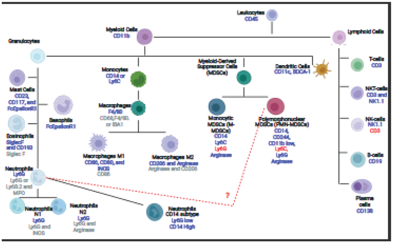
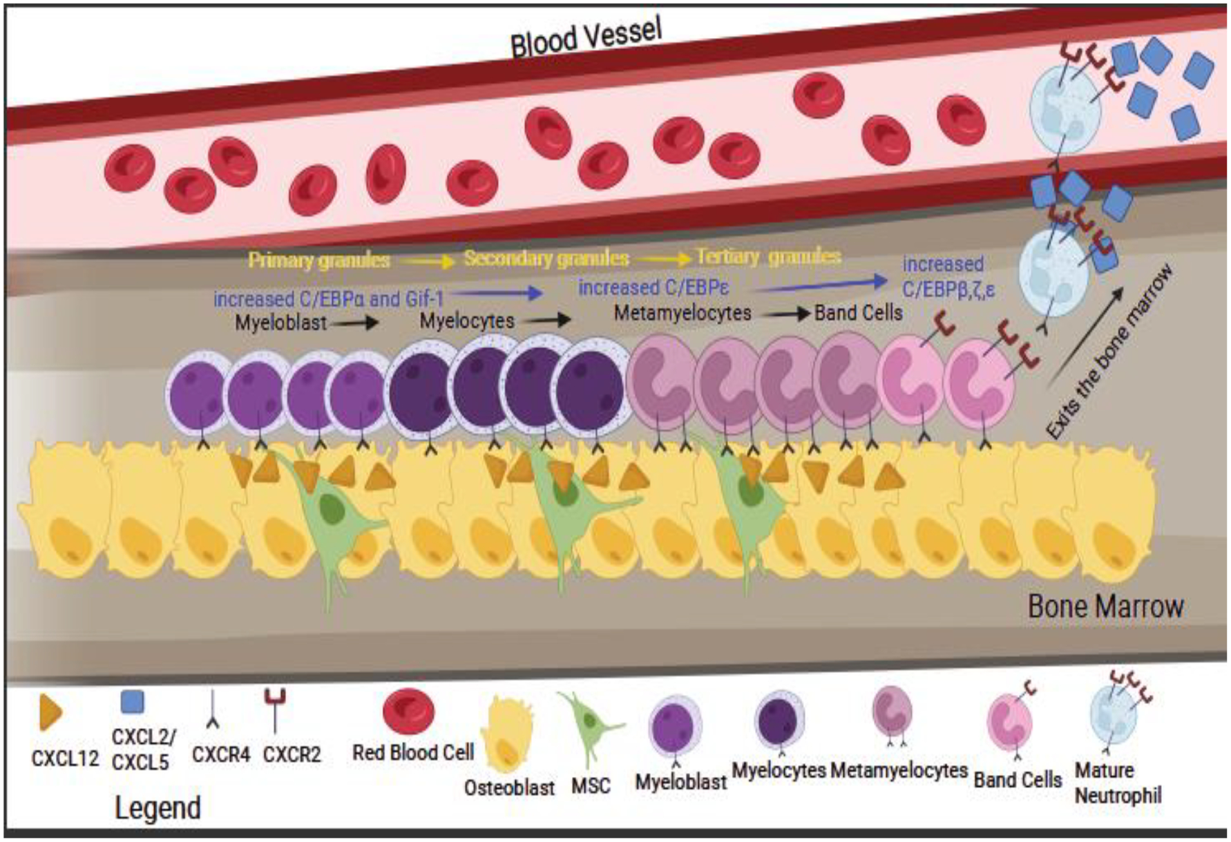
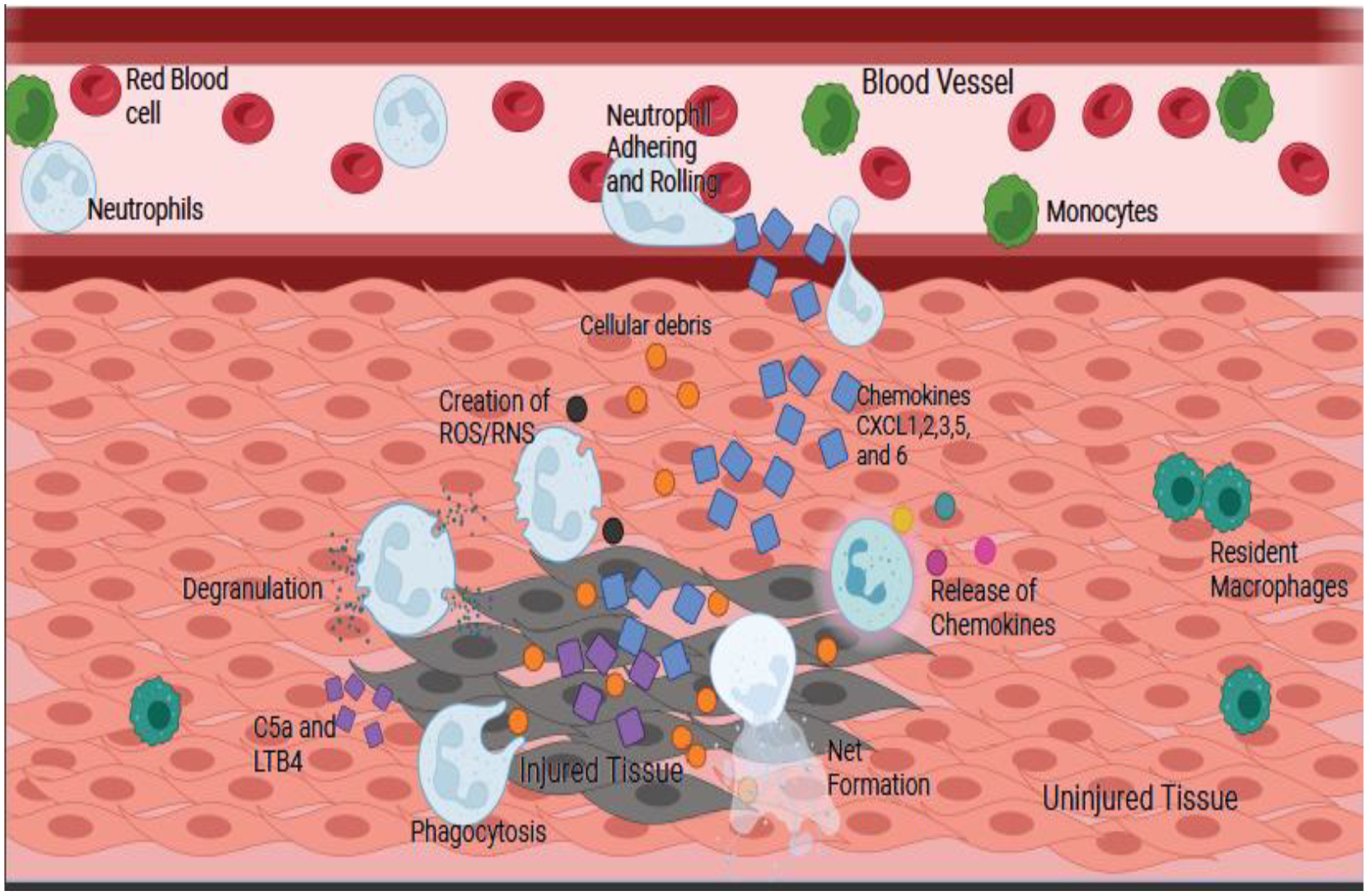
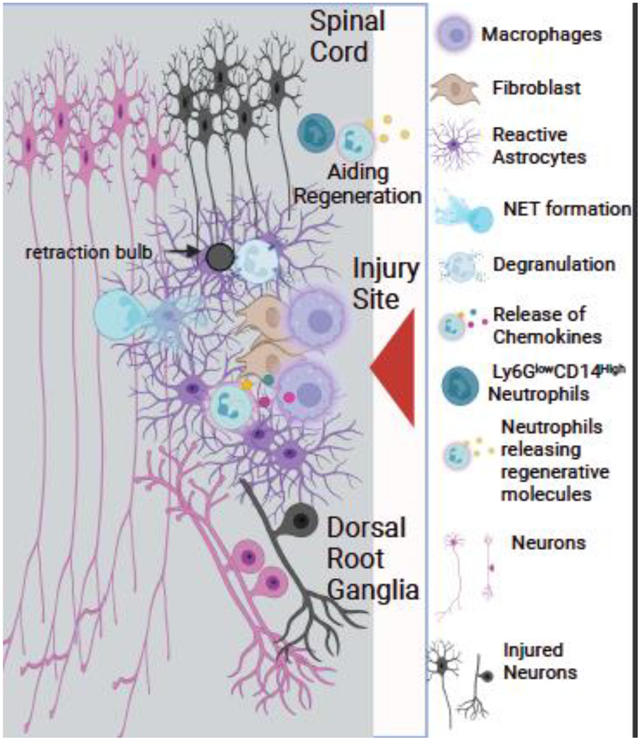
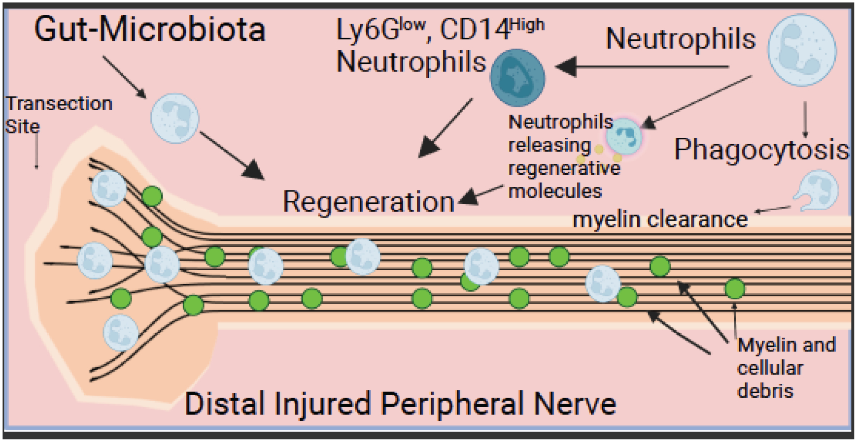
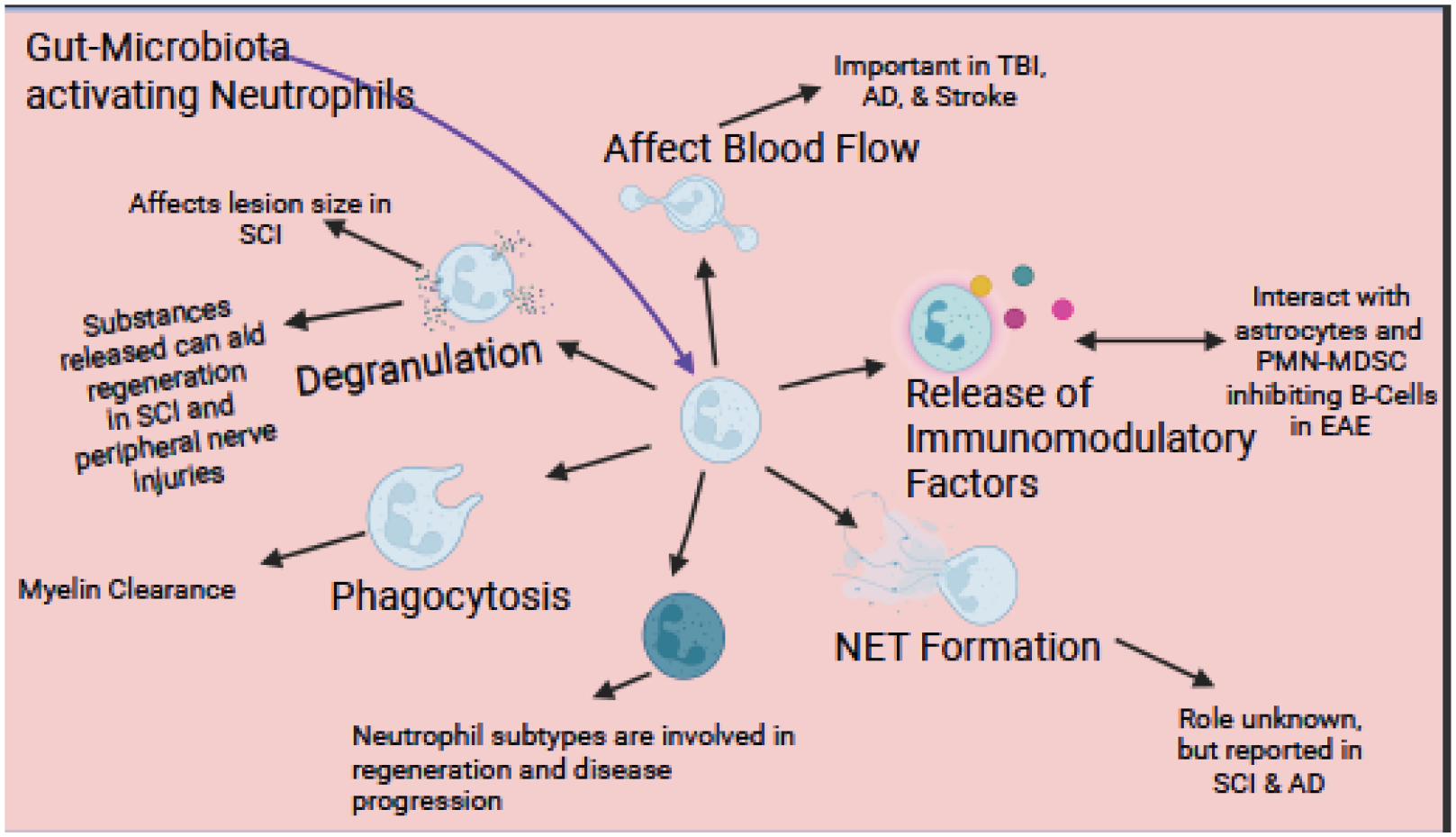
The role of inflammation in nervous system injury and disease is attracting increased attention. Much of that research has focused on microglia in the central nervous system (CNS) and macrophages in the peripheral nervous system (PNS). Much less attention has been paid to the roles played by neutrophils. Neutrophils are part of the granulocyte subtype of myeloid cells. These cells, like macrophages, originate and differentiate in the bone marrow from which they enter the circulation. After tissue damage or infection, neutrophils are the first immune cells to infiltrate into tissues and are directed there by specific chemokines, which act on chemokine receptors on neutrophils. We have reviewed here the basic biology of these cells, including their differentiation, the types of granules they contain, the chemokines that act on them, the subpopulations of neutrophils that exist, and their functions. We also discuss tools available for identification and further study of neutrophils. We then turn to a review of what is known about the role of neutrophils in CNS and PNS diseases and injury, including stroke, Alzheimer's disease, multiple sclerosis, amyotrophic lateral sclerosis, spinal cord and traumatic brain injuries, CNS and PNS axon regeneration, and neuropathic pain. While in the past studies have focused on neutrophils deleterious effects, we will highlight new findings about their benefits. Studies on their actions should lead to identification of ways to modify neutrophil effects to improve health.

Keywords: Chemokine; Nerve injury; Neutrophil; Neutrophil extracellular trap; Phagocytosis; Polymorphonuclear leukocyte; Wallerian degeneration.


Conflict of interest statement

Declaration of Competing Interest The authors have no conflict of interests to disclose.

Figures

Fig. 1.
Fig. 1.. Diagram of Identification Markers to Separate Neutrophils from Basophils, Eosinophils, Mast cells, Macrophages, and Monocytes.
The diagram shows the different cell types within the myeloid and lymphoid lineages. The blue text under each cell type indicates a positive flow marker for that cell type, while the red text indicates a cell marker that is not present in the cell type. The grey text for macrophages, neutrophils, and eosinophils are markers that can be used in immunohistofluorescence to detect these cell types. This list is not an extensive list of cell types as some can be further divided, nor an exhaustive list of cellular markers.
Fig. 2.
Fig. 2.. Development of Neutrophils.
The figure shows the development of neutrophils in the bone marrow from myeloblast to myelocytes, where C/EBPε expression is increased, and primary granules start to form. These cells develop in close contact with osteoblasts and mesenchymal stem cells (MSC), as they produce CXCL12 that binds to the receptor CXCR4 present on the neutrophil precursors. As the cells further develop, they will become metamyelocytes and then band cells with an increase in expression of Gif-1 and C/EBPβ,ζ, and ζ, with secondary and tertiary granules forming. As band cells, the receptor CXCR2 is upregulated, resulting in the mature neutrophils leaving the bone marrow. The yellow text indicates the order of granule formation, the blue text indicates gene expression, and the black text indicates the order of cell differentiation during development.
Fig. 3.
Fig. 3.. Neutrophil Chemotaxis and Mechanism of Action.
Neutrophils will follow the gradient of chemokines to the injury site, then adhere and roll along the vessel before firmly adhering and entering the tissue. Entering the tissue is accomplished by degranulation of the tertiary granules. Once in the tissue, the cytokines CXCL1, 2, 3, 5, 6, LTB4 and C5a, which are released by the injured tissue, will trigger different cellular functions leading to degranulation, phagocytosis, creation of reactive oxygen and nitrogen species (ROS/RNS), formation of NETs, and release of chemokines. The orange circles are cellular debris, the blue squares are chemokines, and the purple squares are C5a and LTB4. The cells are labeled in the figure.
Fig. 4.
Fig. 4.. Neutrophils are Involved in Myelin Clearance.
Wild type and Ccr2 KO mice were pretreated with antibodies against Ly6G to deplete neutrophils or were given an isotope control antibody. The sciatic nerve was then transected unilaterally, and seven days later, the nerve was stained with luxol fast blue (LFB) as an index of intact myelin. The top graph shows the effectiveness of the deletion strategy (A). Graph B shows the percent area stained. The LFB images show residual staining after neutrophil depletion (C-F). (Adapted from Lindborg et al., 2017).
Fig. 5.
Fig. 5.. Neutrophils in Spinal Cord Injury.
After a spinal cord injury (SCI), neutrophils enter the injury site, followed by macrophages. Activated astrocytes keep the neutrophils from spreading in the injury site. Mpo KO mice have a decreased lesion size compared to wild type demonstrating Mpo can have a detrimental effect. Several molecules released by neutrophils are beneficial to regeneration. Additionally, the neutrophil Ly6GlowCD14+ subtype has been shown to aid regeneration. Darkened neurons (motor and sensory) indicate injured neurons.
Fig. 6.
Fig. 6.. Neutrophils in Peripheral Nerve Injuries.
After peripheral nerve injury, the distal nerve undergoes Wallerian degeneration (WD). Neutrophils play a role in WD, as depleting neutrophils delays myelin clearance. Many molecules (i.e., oncomodulin, nerve growth factor, & neutrophil peptide-1) aid regeneration that are released from neutrophils. Changes in the gut microbiota have been shown to accelerate regeneration, which is dependent on neutrophils. Additionally, the Ly6GlowCD14+ subtype is involved in regeneration after a peripheral nerve injury.
Fig. 7.
Fig. 7.. Summary.
In this figure, we summarize all the effects discussed in this article. Neutrophils have many vital roles in neurological disease and injuries. Degranulation affects the lesion size in multiple sclerosis (MS) and spinal cord injuries (SCI). Neutrophils possibly help with the phagocytosis of myelin debris after injury. The CD14 subtype’s role in regeneration is just starting to be investigated but holds promise. Other subtypes; (i.e., N2) involvement in other neurological conditions is being investigated. NET formation has been reported in SCI and Alzheimer’s disease (AD). Cytokine release from neutrophils can activate other cell types causing a continued immune response. Neutrophils have a significant effect on blood flow which affects clinical outcomes during traumatic brain injury (TBI) and stroke, in addition to playing a role in AD. It has been shown that neutrophils release regenerative molecules that aid regeneration in the peripheral nervous system after injury and in SCI. The role of the gut-microbiota activating neutrophils to aid in regeneration is an exciting and new avenue of research.

Similar articles

Cited by

References

    1. Abdel-Latif D, Steward M, Macdonald DL, Francis GA, Dinauer MC, Lacy P, 2004. Rac2 is critical for neutrophil primary granule exocytosis. Blood 104, 832–839. 10.1182/blood-2003-07-2624 - DOI - PubMed
    1. Ahuja SK, Lee JC, Murphy PM, 1996. CXC chemokines bind to unique sets of selectivity determinants that can function independently and are broadly distributed on multiple domains of human interleukin-8 receptor B. Determinants of high affinity binding and receptor activation are distinct. J Biol Chem 271, 225–232. 10.1074/jbc.271.1.225 - DOI - PubMed
    1. Ahuja SK, Murphy PM, 1996. The CXC chemokines growth-regulated oncogene (GRO) alpha, GRObeta, GROgamma, neutrophil-activating peptide-2, and epithelial cell-derived neutrophil-activating peptide-78 are potent agonists for the type B, but not the type A, human interleukin-8 receptor. J Biol Chem 271, 20545–20550. 10.1074/jbc.271.34.20545 - DOI - PubMed
    1. Allport JR, Ding H, Collins T, Gerritsen ME, Luscinskas FW, 1997. Endothelial-dependent mechanisms regulate leukocyte transmigration: a process involving the proteasome and disruption of the vascular endothelial-cadherin complex at endothelial cell-to-cell junctions. J Exp Med 186, 517–527. 10.1084/jem.186.4.517 - DOI - PMC - PubMed
    1. Amaral FA, Costa VV, Tavares LD, Sachs D, Coelho FM, Fagundes CT, Soriani FM, Silveira TN, Cunha LD, Zamboni DS, Quesniaux V, Peres RS, Cunha TM, Cunha FQ, Ryffel B, Souza DG, Teixeira MM, 2012. NLRP3 inflammasome-mediated neutrophil recruitment and hypernociception depend on leukotriene B4 in a murine model of gout. Arthritis Rheum 64, 474–484. 10.1002/art.33355 - DOI - PubMed

Publication types