

RNA folding pathways from all-atom simulations with a variationally improved history-dependent bias
- PMID: 37355771
- PMCID: PMC10432211
- DOI: 10.1016/j.bpj.2023.06.012
RNA folding pathways from all-atom simulations with a variationally improved history-dependent bias
Abstract
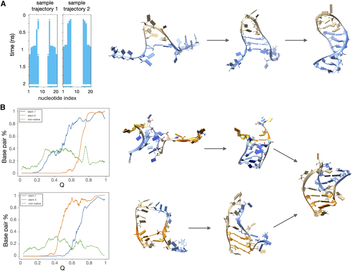
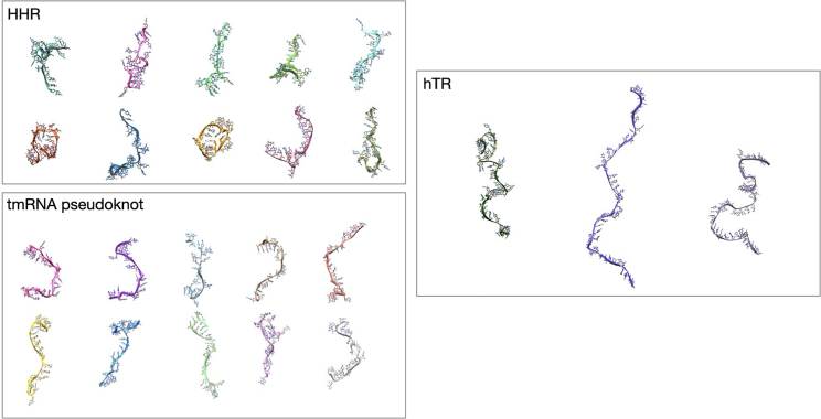
Atomically detailed simulations of RNA folding have proven very challenging in view of the difficulties of developing realistic force fields and the intrinsic computational complexity of sampling rare conformational transitions. As a step forward in tackling these issues, we extend to RNA an enhanced path-sampling method previously successfully applied to proteins. In this scheme, the information about the RNA's native structure is harnessed by a soft history-dependent biasing force promoting the generation of productive folding trajectories in an all-atom force field with explicit solvent. A rigorous variational principle is then applied to minimize the effect of the bias. Here, we report on an application of this method to RNA molecules from 20 to 47 nucleotides long and increasing topological complexity. By comparison with analog simulations performed on small proteins with similar size and architecture, we show that the RNA folding landscape is significantly more frustrated, even for relatively small chains with a simple topology. The predicted RNA folding mechanisms are found to be consistent with the available experiments and some of the existing coarse-grained models. Due to its computational performance, this scheme provides a promising platform to efficiently gather atomistic RNA folding trajectories, thus retain the information about the chemical composition of the sequence.
Copyright © 2023 Biophysical Society. Published by Elsevier Inc. All rights reserved.
Conflict of interest statement
Declaration of interests P.F. is cofounder of Sibylla Biotech (www.sibyllabiotech.it), a company involved in early-stage drug discovery.
Figures


Similar articles
-
Variational scheme to compute protein reaction pathways using atomistic force fields with explicit solvent.Phys Rev Lett. 2015 Mar 6;114(9):098103. doi: 10.1103/PhysRevLett.114.098103. Epub 2015 Mar 4. Phys Rev Lett. 2015. PMID: 25793854
-
A critical comparison of coarse-grained structure-based approaches and atomic models of protein folding.Phys Chem Chem Phys. 2017 May 31;19(21):13629-13639. doi: 10.1039/c7cp01532a. Phys Chem Chem Phys. 2017. PMID: 28530269
-
Computing Reaction Pathways of Rare Biomolecular Transitions using Atomistic Force-Fields.Biophys Chem. 2016 Jan;208:62-7. doi: 10.1016/j.bpc.2015.06.014. Epub 2015 Aug 14. Biophys Chem. 2016. PMID: 26320390 Review.
-
Multiscale investigation of chemical interference in proteins.J Chem Phys. 2010 May 7;132(17):175101. doi: 10.1063/1.3404401. J Chem Phys. 2010. PMID: 20459186
-
Atomistic molecular simulations of protein folding.Curr Opin Struct Biol. 2012 Feb;22(1):52-61. doi: 10.1016/j.sbi.2011.12.001. Epub 2012 Jan 17. Curr Opin Struct Biol. 2012. PMID: 22257762 Review.
Cited by
-
Computer-aided comprehensive explorations of RNA structural polymorphism through complementary simulation methods.QRB Discov. 2022 Oct 17;3:e21. doi: 10.1017/qrd.2022.19. eCollection 2022. QRB Discov. 2022. PMID: 37529277 Free PMC article. Review.
References
-
- Sharp P.A. The Centrality of RNA. Cell. 2009;136:577–580. https://www.cell.com/cell/abstract/S0092-8674(09)00143-3 - PubMed
-
- Balvay L., Lopez Lastra M., et al. Ohlmann T. Translational control of retroviruses. Nat. Rev. Microbiol. 2007;5:128–140. https://www.nature.com/articles/nrmicro1599 - PMC - PubMed
Publication types
MeSH terms
Substances
LinkOut - more resources
Full Text Sources
Research Materials
