Longitudinal Patterns of Brain Changes in a Community Sample in Relation to Aging and Cognitive Status
- PMID: 37355895
- PMCID: PMC10674101
- DOI: 10.3233/JAD-230080
Longitudinal Patterns of Brain Changes in a Community Sample in Relation to Aging and Cognitive Status
Abstract
Background: Aging and Alzheimer's disease (AD) are characterized by widespread cortical and subcortical atrophy. Though atrophy patterns between aging and AD overlap considerably, regional differences between these two conditions may exist. Few studies, however, have investigated these patterns in large community samples.
Objective: Elaborate longitudinal changes in brain morphometry in relation to aging and cognitive status in a well-characterized community cohort.
Methods: Clinical and neuroimaging data were compiled from 72 participants from the Cardiovascular Health Study-Cognition Study, a community cohort of healthy aging and probable AD participants. Two time points were identified for each participant with a mean follow-up time of 5.36 years. MRI post-processing, morphometric measurements, and statistical analyses were performed using FreeSurfer, Version 7.1.1.
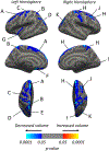
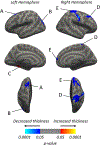
Results: Cortical volume was significantly decreased in the bilateral superior frontal, bilateral inferior parietal, and left superior parietal regions, among others. Cortical thickness was significantly reduced in the bilateral superior frontal and left inferior parietal regions, among others. Overall gray and white matter volumes and hippocampal subfields also demonstrated significant reductions. Cortical volume atrophy trajectories between cognitively stable and cognitively declined participants were significantly different in the right postcentral region.
Conclusion: Observed volume reductions were consistent with previous studies investigating morphometric brain changes. Patterns of brain atrophy between AD and aging may be different in magnitude but exhibit widespread spatial overlap. These findings help characterize patterns of brain atrophy that may reflect the general population. Larger studies may more definitively establish population norms of aging and AD-related neuroimaging changes.
Keywords: Aging; Alzheimer’s disease; cognition; cognitive dysfunction; neuroimaging.
Conflict of interest statement
CONFLICT OF INTEREST
Dr. Raji is an Editorial Board Member of this journal but was not involved in the peer-review process nor had access to any information regarding its peerreview. Dr. Raji does consult to Brainreader ApS, Apollo Health, Neurevolution LLC, and the Pacific Neuroscience Foundation.
Figures


Similar articles
-
Patterns of Grey Matter Atrophy at Different Stages of Parkinson's and Alzheimer's Diseases and Relation to Cognition.Brain Topogr. 2019 Jan;32(1):142-160. doi: 10.1007/s10548-018-0675-2. Epub 2018 Sep 11. Brain Topogr. 2019. PMID: 30206799
-
Structural neuroimaging changes associated with subjective cognitive decline from a clinical sample.Neuroimage Clin. 2024;42:103615. doi: 10.1016/j.nicl.2024.103615. Epub 2024 May 10. Neuroimage Clin. 2024. PMID: 38749146 Free PMC article.
-
Five-Year Longitudinal Brain Volume Change in Healthy Elders at Genetic Risk for Alzheimer's Disease.J Alzheimers Dis. 2017;55(4):1363-1377. doi: 10.3233/JAD-160504. J Alzheimers Dis. 2017. PMID: 27834774 Free PMC article.
-
Modeling grey matter atrophy as a function of time, aging or cognitive decline show different anatomical patterns in Alzheimer's disease.Neuroimage Clin. 2019;22:101786. doi: 10.1016/j.nicl.2019.101786. Epub 2019 Mar 19. Neuroimage Clin. 2019. PMID: 30921610 Free PMC article.
-
Structural magnetic resonance imaging for the early diagnosis of dementia due to Alzheimer's disease in people with mild cognitive impairment.Cochrane Database Syst Rev. 2020 Mar 2;3(3):CD009628. doi: 10.1002/14651858.CD009628.pub2. Cochrane Database Syst Rev. 2020. PMID: 32119112 Free PMC article.
Cited by
-
Automated brain segmentation and volumetry in dementia diagnostics: a narrative review with emphasis on FreeSurfer.Front Aging Neurosci. 2024 Sep 3;16:1459652. doi: 10.3389/fnagi.2024.1459652. eCollection 2024. Front Aging Neurosci. 2024. PMID: 39291276 Free PMC article. Review.
References
-
- Conde JR, Streit WJ (2006) Microglia in the aging brain. J Neuropathol Exp Neurol 65, 199–203. - PubMed
-
- Rutten BP, Schmitz C, Gerlach OH, Oyen HM, de Mesquita EB, Steinbusch HW, Korr H (2007) The aging brain: Accumulation of DNA damage or neuron loss? Neurobiol Aging 28, 91–98. - PubMed
-
- Morrison JH, Hof PR (2007) Life and death of neurons in the aging cerebral cortex. Int Rev Neurobiol 81, 41–57. - PubMed
-
- Khachaturian ZS (1985) Diagnosis of Alzheimer’s disease. Arch Neurol 42, 1097–1105. - PubMed
Publication types
MeSH terms
Grants and funding
- N01 HC085080/HL/NHLBI NIH HHS/United States
- U01 HL080295/HL/NHLBI NIH HHS/United States
- N01 HC085082/HL/NHLBI NIH HHS/United States
- U01 HL130114/HL/NHLBI NIH HHS/United States
- N01 HC055222/HL/NHLBI NIH HHS/United States
- N01 HC085079/HL/NHLBI NIH HHS/United States
- 75N92021D00006/HL/NHLBI NIH HHS/United States
- R01 AG023629/AG/NIA NIH HHS/United States
- N01 HC085081/HL/NHLBI NIH HHS/United States
- HHSN268200800007C/HL/NHLBI NIH HHS/United States
- N01 HC085086/HL/NHLBI NIH HHS/United States
- N01 HC085083/HL/NHLBI NIH HHS/United States
- HHSN268201200036C/HL/NHLBI NIH HHS/United States
- HHSN268201800001C/HL/NHLBI NIH HHS/United States
LinkOut - more resources
Full Text Sources
Medical

