Review
. 2024 Mar;34(2):e13184.
doi: 10.1111/bpa.13184. Epub 2023 Jun 25.

Pathology explains various mechanisms of auto-immune inflammatory peripheral neuropathies

Affiliations
Review

Pathology explains various mechanisms of auto-immune inflammatory peripheral neuropathies

Jean-Michel Vallat et al. Brain Pathol. 2024 Mar.

Abstract

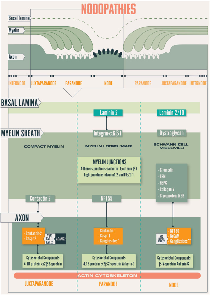
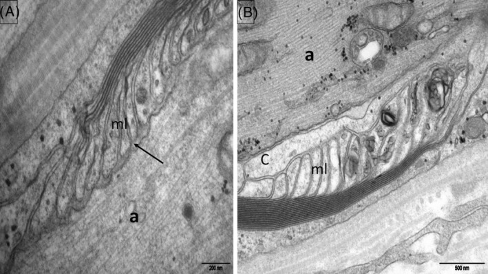
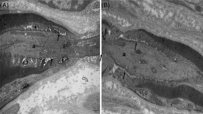
Autoimmune neuropathies are a heterogeneous group of rare and disabling diseases in which the immune system is thought to target antigens in the peripheral nervous system: they usually respond to immune therapies. Guillain-Barré syndrome is divided into several subtypes including "acute inflammatory demyelinating polyradiculoneuropathy," "acute motor axonal neuropathy," "acute motor sensory neuropathy," and other variants. Chronic forms such as chronic inflammatory demyelinating polyneuropathy (CIDP) and other subtypes and polyneuropathy associated with IgM monoclonal gammopathy; autoimmune nodopathies also belong to this group of auto-immune neuropathies. It has been shown that immunoglobulin G from the serum of about 30% of CIDP patients immunolabels nodes of Ranvier or paranodes of myelinated axons. Whatever the cause of myelin damage of the peripheral nervous system, the initial attack on myelin by a dysimmune process may begin either at the internodal area or in the paranodal and nodal regions. The term "nodoparanodopathy" was first applied to some "axonal Guillain-Barré syndrome" subtypes, then extended to cases classified as CIDP bearing IgG4 antibodies against paranodal axoglial proteins. In these cases, paranodal dissection develops in the absence of macrophage-induced demyelination. In contrast, the mechanisms of demyelination of other dysimmune neuropathies induced by macrophages are unexplained, as no antibodies have been identified in such cases. The main objective of this presentation is to show that the pathology illustrates, confirms, and may explain such mechanisms.

Keywords: internode; myelin; neurofascin; node; nodoparanodopathy; paranode.


Conflict of interest statement

The authors declare no conflict of interest.

Figures

FIGURE 1
FIGURE 1
Schematic representation of the node of Ranvier. Caspr, contactin‐associated protein; ERM, Ezrin‐Radixin‐Moesin; KCNQ, potassium channel, voltage‐gated, KQT‐like subfamily Kv, potassium channel; MAG, myelin‐associated glycoprotein; Nav, sodium channel; NF155, neurofascin‐155; NF186, neurofascin‐186/140; VGC, voltage‐gated channel.
FIGURE 2
FIGURE 2
Normal control subject. Electron micrographs, longitudinal sections of the nodoparanodal area (sural nerve biopsy). (A) Paranode, node, microvilli (mv), axon (a). (B) Myelin (ml) loops are attached to the axolemma by periodic, punctiform, dense structures corresponding to the transverse bands (arrow).
FIGURE 3
FIGURE 3
Classical CIDP. Paraffin‐embedded longitudinal section (sural nerve biopsy) with anti‐CD3 antibody staining. A vessel of the perineurium (P) is surrounded by T‐lymphocytes and a few are also scattered in the endoneurium (arrows). CIDP, chronic inflammatory demyelinating polyneuropathy.
FIGURE 4
FIGURE 4
Classical CIDP. Electron micrograph, longitudinal section (sural nerve biopsy). In an internodal domain several macrophages (M) loaded with myelin debris are in contact to a large diameter axon (Ax) completely devoid of myelin; they are located between the axon and the Schwann cell basal lamina (arrow). Some other macrophages (m) are identified in the interstitial tissue. CIDP, chronic inflammatory demyelinating polyneuropathy.
FIGURE 5
FIGURE 5
Anti‐NF155 paranodopathy. Electron micrograph, longitudinal section (sural nerve biopsy). (A): The transverse bands between the myelin loops (ml) and the axon (a) are absent (arrow). (B): A cellular process (C) is interposed between the myelin loops (ml) and the axon (a).
FIGURE 6
FIGURE 6
Anti‐Caspr1 (A) and anti‐pan‐neurofascin (B) paranodopathies. Electron micrographs, longitudinal sections (sural nerve biopsy). Most transverse bands are missing (arrows); myelin loops (ml) are retracted and narrow (a: axon).

Similar articles

Cited by

References

    1. Lehmann HC, Wunderlich G, Fink GR, Sommer C. Diagnosis of peripheral neuropathy. Neurol Res Pract. 2020;2:20. 10.1186/s42466-020-00064-2 - DOI - PMC - PubMed
    1. Salzer JL. Schwann cell myelination. Cold Spring Harb Perspect Biol. 2015;7(8):a020529. 10.1101/cshperspect.a020529 - DOI - PMC - PubMed
    1. Uncini A, Susuki K, Yuki N. Nodo‐paranodopathy: beyond the demyelinating and axonal classification in anti‐ganglioside antibody‐mediated neuropathies. Clin Neurophysiol. 2013;124(10):1928–34. 10.1016/j.clinph.2013.03.025 - DOI - PubMed
    1. Uncini A, Vallat JM. Autoimmune nodo‐paranodopathies of peripheral nerve: the concept is gaining ground. J Neurol Neurosurg Psychiatry. 2018;89(6):627–35. 10.1136/jnnp-2017-317192 - DOI - PubMed
    1. Devaux JJ, Odaka M, Yuki N. Nodal proteins are target antigens in Guillain–Barré syndrome. J Peripher Nerv Syst. 2012;17(1):62–71. 10.1111/j.1529-8027.2012.00372.x - DOI - PubMed

MeSH terms