. 2023 Jun 25;13(1):10296.
doi: 10.1038/s41598-023-37475-5.

A predictive signal model for dynamic cardiac magnetic resonance imaging

Affiliations

A predictive signal model for dynamic cardiac magnetic resonance imaging

Aaron D Curtis et al. Sci Rep. .

Abstract

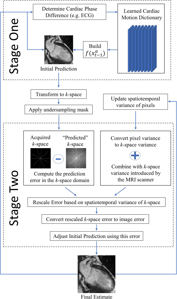
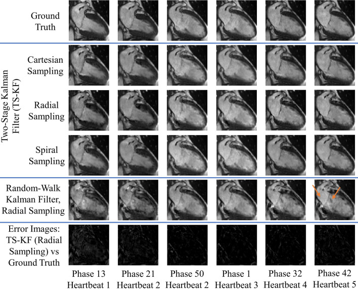
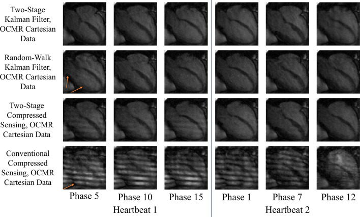
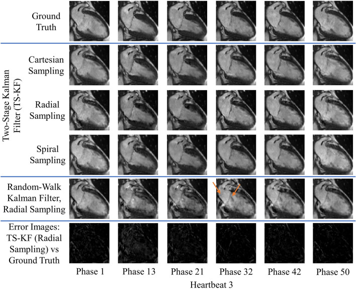
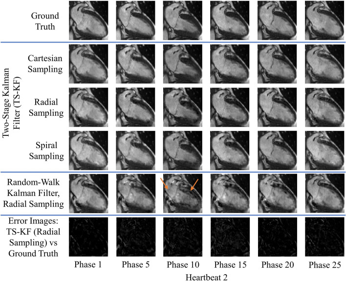
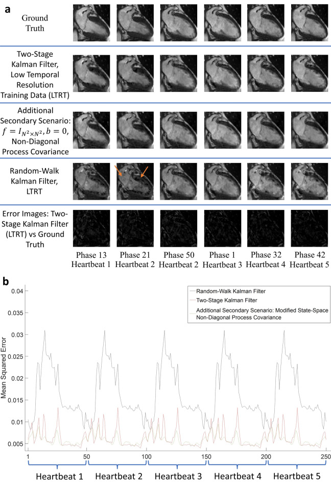
Robust dynamic cardiac magnetic resonance imaging (MRI) has been a long-standing endeavor-as real-time imaging can provide information on the temporal signatures of disease we currently cannot assess-with the past decade seeing remarkable advances in acceleration using compressed sensing (CS) and artificial intelligence (AI). However, substantial limitations to real-time imaging remain and reconstruction quality is not always guaranteed. To improve reconstruction fidelity in dynamic cardiac MRI, we propose a novel predictive signal model that uses a priori statistics to adaptively predict temporal cardiac dynamics. By using a small training set obtained from the same patient, the new signal model can achieve robust dynamic cardiac MRI in the presence of irregular cardiac rhythm. Evaluation on simulated irregular cardiac dynamics and prospectively undersampled clinical cardiac MRI data demonstrate improved reconstruction quality for two reconstruction frameworks: Kalman filter and CS. The predictive model also works with different undersampling patterns (cartesian, radial, spiral) and can serve as a versatile foundation for robust dynamic cardiac MRI.


Conflict of interest statement

The authors declare no competing interests.

Figures

Figure 1
Figure 1
A predictive signal model incorporated in a two-stage Kalman filter. Stage One uses a learned cardiac motion dictionary to predict the next cardiac image. Stage Two uses the spatiotemporal statistics and the acquired k-space data to update this prediction and produce the final estimate.
Figure 2
Figure 2
Reconstruction of multiple cardiac cycles I. These reconstructions were performed on the first UK Biobank dataset. The orange arrows illustrate distortions in the shape of the left atrium and the contrast along the wall of the left ventricle during random-walk Kalman filter reconstructions. Our two-stage Kalman filter demonstrates improved tracking and reconstruction quality compared to the random-walk Kalman filter.
Figure 3
Figure 3
Reconstruction of multiple cardiac cycles II. These reconstructions were performed on the prospectively undersampled OCMR dataset. The orange arrows illustrate the inability of the random-walk Kalman filter and conventional CS to properly model cardiac dynamics for the given undersampling scheme. Equipped with a predictive signal model, both our two-stage Kalman filter and two-stage CS scheme demonstrate improved tracking and reconstruction quality compared to the random-walk Kalman filter and conventional CS when applied to raw k-space datasets.
Figure 4
Figure 4
Reconstruction of a single arrhythmic event. These reconstructions were performed on the first UK Biobank dataset. Arrhythmia is simulated as skipping phases 8 and 9 in heartbeat 2 and a sudden return to systole (phase 1) in heartbeat 3. The orange arrows illustrate distortions in the shape of the left atrium and the contrast along the wall of the left ventricle during random-walk Kalman filter reconstructions. The two-stage Kalman filter can track the arrhythmic event and continuously provide high-quality reconstructions afterwards.
Figure 5
Figure 5
Reconstruction in the presence of variable sinus rhythm. These reconstructions were performed on the first UK Biobank dataset. A doubling of the sinus rhythm is simulated by skipping every other phase in heartbeat 2. The orange arrows demonstrate perturbations in the shape of the left atrium and the contrast along the wall of the left ventricle during random-walk Kalman filter reconstructions. Our two-stage Kalman filter demonstrates improved tracking and reconstruction quality compared to the random-walk Kalman filter.
Figure 6
Figure 6
Mean-squared error (MSE) plots for the second primary scenario (single arrhythmic event), and MSE statistics for all scenarios. The subplots demonstrate the MSE for the second primary scenario across all UK Biobank datasets: (a) two-stage Kalman filter (TS-KF) with radial sampling, (b) TS-KF with cartesian sampling, (c) random-walk Kalman filter (RW-KF) with radial sampling, and (d) RW-KF with cartesian sampling. The x indices represent the frame number. Except for the index labeled “1”, each index also indicates the last phase of the corresponding cardiac cycle. (a–d) All demonstrate the robustness and consistency of our two-stage Kalman filter. Across all datasets, the MSE exhibited convergence. Subplot (e) demonstrates the maximum, mean, and median (med) values for all primary scenarios using radial sampling across all UK Biobank datasets. The MSE for the OCMR dataset is not available, as prospectively undersampling precludes the availability of the ground truth. Note that in general the TS-KF offered improved or comparable performance to the RW-KF. Subplot (f) demonstrates the maximum, mean, and median values for all primary scenarios using cartesian sampling across all datasets. Note that the TS-KF vastly outperforms the RW-KF for this sampling scheme.
Figure 7
Figure 7
Reconstruction accommodating for low temporal resolution training data. All reconstruction results are shown in (a). The test set has a temporal resolution five times greater than that of the training set. Combined with a spatial undersampling factor of 12.5, the effective acceleration factor is 100.5. Note that the random-walk Kalman filter reconstructs erroneous images: this is a consequence of inaccurate modelling. To compensate, the Kalman filter continuously attempts to reconstruct the mean, hence the perturbations. These perturbations are highlighted with orange arrows. Subplot (b) provides the MSE for the reconstructions shown in (a). The MSE plot for our two-stage Kalman filter simulation is shown in red. Our algorithm properly accommodates low temporal resolution training data by ensuring convergent behavior. Furthermore, the magnitude of the error is comparable to those shown in Fig. 6.

References

    1. Ghugre NR, Pop M, Barry J, Connelly KA, Wright GA. Quantitative magnetic resonance imaging can distinguish remodeling mechanisms after acute myocardial infarction based on the severity of ischemic insult. Magn. Reson. Med. 2013 doi: 10.1002/mrm.24531. - DOI - PubMed
    1. Tseng W-YI, Su M-YM, Tseng Y-HE. Introduction to cardiovascular magnetic resonance: Technical principles and clinical applications. Acta Cardiol. Sin. 2016;32:129–144. - PMC - PubMed
    1. Uecker M, et al. Real-time MRI at a resolution of 20 ms. NMR Biomed. 2010 doi: 10.1002/nbm.1585. - DOI - PubMed
    1. Kellman P, et al. High spatial and temporal resolution cardiac cine MRI from retrospective reconstruction of data acquired in real time using motion correction and resorting. Magn. Reson. Med. 2009 doi: 10.1002/mrm.22153. - DOI - PMC - PubMed
    1. Xue H, Kellman P, Larocca G, Arai AE, Hansen MS. High spatial and temporal resolution retrospective cine cardiovascular magnetic resonance from shortened free breathing real-time acquisitions. J. Cardiovasc. Magn. Reson. 2013;15:102. doi: 10.1186/1532-429X-15-102. - DOI - PMC - PubMed

Publication types

Grants and funding