. 2023 Jun 25;14(1):168.
doi: 10.1186/s13287-023-03396-3.

Mesenchymal progenitor cells from non-inflamed versus inflamed synovium post-ACL injury present with distinct phenotypes and cartilage regeneration capacity

Affiliations

Mesenchymal progenitor cells from non-inflamed versus inflamed synovium post-ACL injury present with distinct phenotypes and cartilage regeneration capacity

Roman J Krawetz et al. Stem Cell Res Ther. .

Abstract

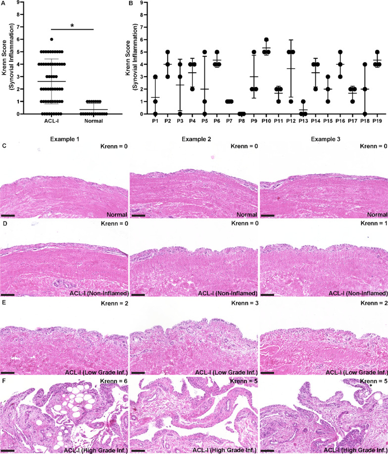
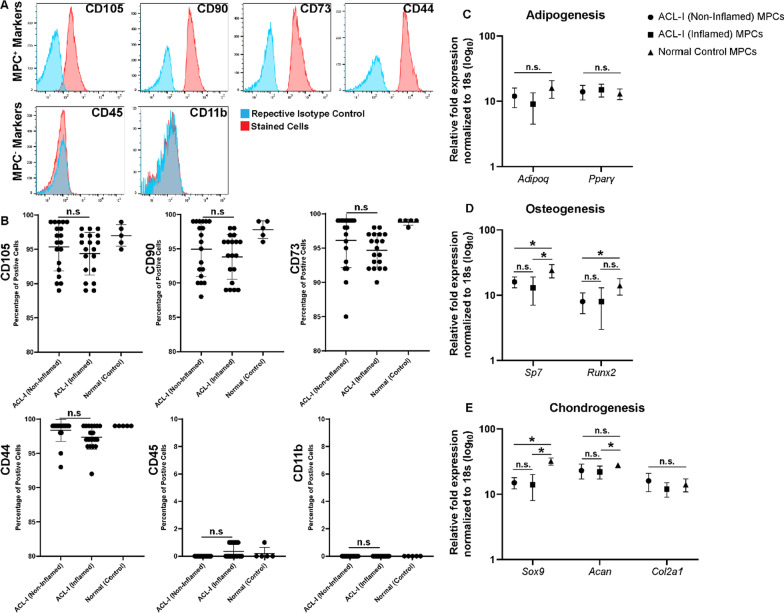
Background: Osteoarthritis (OA) is a chronic debilitating disease impacting a significant percentage of the global population. While there are numerous surgical and non-invasive interventions that can postpone joint replacement, there are no current treatments which can reverse the joint damage occurring during the pathogenesis of the disease. While many groups are investigating the use of stem cell therapies in the treatment of OA, we still don't have a clear understanding of the role of these cells in the body, including heterogeneity of tissue resident adult mesenchymal progenitor cells (MPCs).

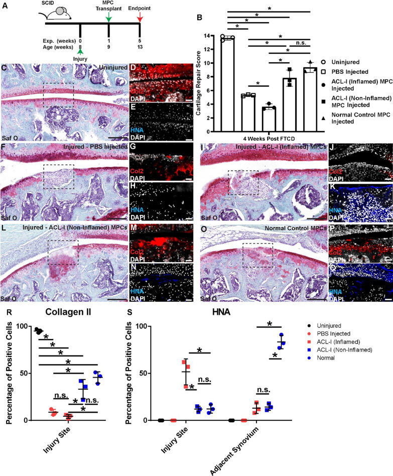
Methods: In the current study, we examined MPCs from the synovium and individuals with or without a traumatic knee joint injury and explored the chondrogenic differentiation capacity of these MPCs in vitro and in vivo.

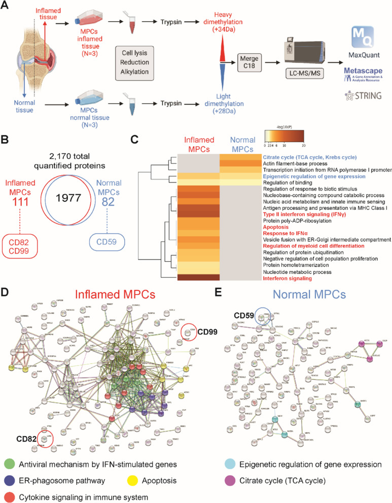
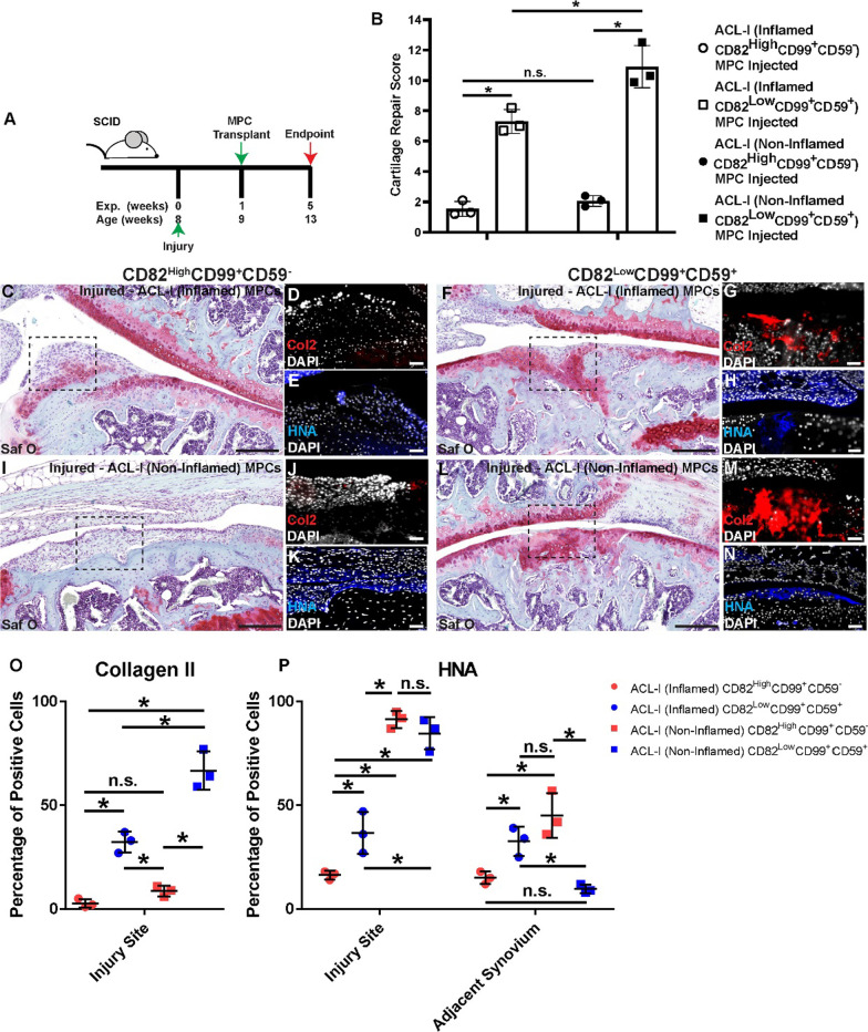
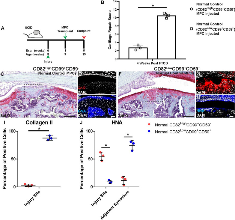
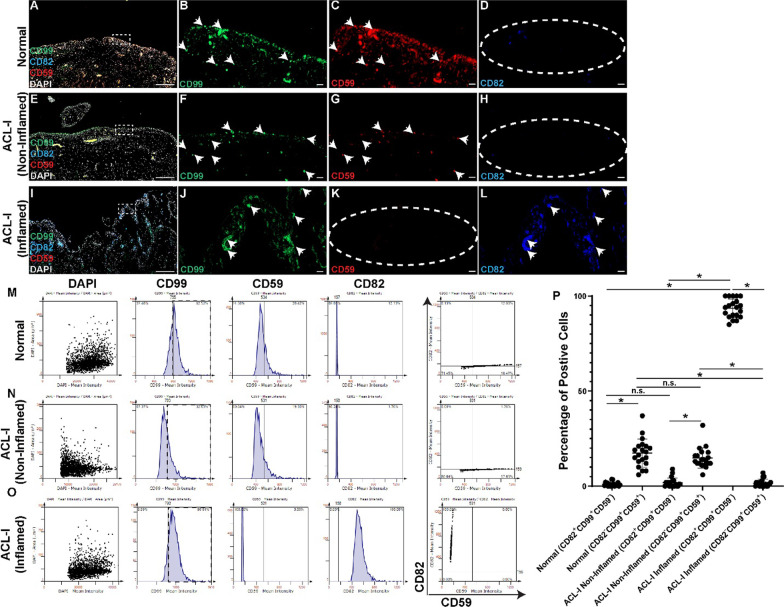
Results: We found that there is heterogeneity of MPCs with the adult synovium and distinct sub-populations of MPCs and the abundancy of these sub-populations change with joint injury. Furthermore, only some of these sub-populations have the ability to effect cartilage repair in vivo. Using an unbiased proteomics approach, we were able to identify cell surface markers that identify this pro-chondrogenic MPC population in normal and injured joints, specifically CD82LowCD59+ synovial MPCs have robust cartilage regenerative properties in vivo.

Conclusions: The results of this study clearly show that cells within the adult human joint can impact cartilage repair and that these sub-populations exist within joints that have undergone a traumatic joint injury. Therefore, these populations can be exploited for the treatment of cartilage injuries and OA in future clinical trials.

Keywords: ACL injury; Inflammation; Mesenchymal progenitor cells; Osteoarthritis.


Conflict of interest statement

Authors declare that they have no competing interests.

Figures

Fig. 1
Fig. 1
Synovitis within ACL-I and normal joints. Synovitis was graded within ACL-I (n = 19, 3 biopsies per individual) and normal knee joints (n = 5, 3 biopsies per individual) using the histological grading score developed by Krenn et al. (A). Individual biopsies were graded within every joint and heterogeneity was observed between biopsies locations within a given joints and also between patients (B). Three representative images of normal (C) and ACL-I (DF) synovium presenting with no, low-, and high-grade synovitis. Scale bar equal 100 µm. *p < 0.05
Fig. 2
Fig. 2
Phenotypic characterization of synovial MPCs. Flow cytometry was employed to quantify the expression of the MPC+ (CD105, CD90, CD73, CD44) and MPC (CD45, CD11b) cell surface markers (A). The relative expression of each marker was quantified within each group and compared between groups (B). No differences in any marker were observed between ACL-I groups. Adipogenic (C), osteogenic (D) and chondrogenic (E) differentiation was assayed by qPCR expression using lineage specific markers. n.s. = not significant; *p < 0.05
Fig. 3
Fig. 3
Transplanted human MPCs into mice with a full thickness cartilage defect. Synovial MPCs were injected in NGS mice post-FTCD (A). Cartilage repair across all groups was quantified (B). Safranin O staining of uninjured (C), injured mice injected with saline (F) or MPCs from ACL-I (Inflamed, n = 3 patients) (I), ACL-I (Non-Inflamed, n = 3 patients) (L) or normal (n = 3 three patients) (O) synovium. Col2 (red: D, G, J, M, P) and HNA (blue: E, H, K, N, Q) staining within each group is also presented. Note: The cell lines injected into mice shown in images I and L were derived from the same patient, but different biopsies. Tissue cytometry was employed to quantify the number of Collagen 2 positive cells in the injury site (R) and the HNA positive cells within the injury site and adjacent synovium (S). Scale bar equals 75 µm. n.s. = not significant; *p < 0.05
Fig. 4
Fig. 4
Proteomics analysis of human MPC populations from ACL-I joints. Schematic representation of the quantitative shotgun proteomics workflow (A). Distribution of proteins identified in each population (B). Heat map showing pathways regulated by differentially expressed proteins in MPCs from inflamed versus normal synovium generated with Metascape (C). STRING-db analysis of protein–protein interaction networks with elevated proteins in MPCs from inflamed (D) or normal (E) synovium. Colored lines between the proteins indicate different types of interaction evidence: curated databases (teal); experimentally determined interactions (pink); predicted interactions gene neighborhood (green); gene fusions (red); gene co-occurrence (blue); text-mining (yellow); co-expression (black); protein homology (purple). MPC lines derived from n = 3 patients
Fig. 5
Fig. 5
Transplanted sub-populations of human MPCs. Synovial MPCs from ACL-I patients (both inflamed (N = 3) and non-inflamed (N = 3)) were purified based on the expression of CD82 and CD59 and injected in NGS mice with a FTCD (A). Cartilage repair across all groups with CD82HighCD99+CD59 or CD82LowCD99+CD59+ MPCs were quantified (B). Safranin O staining of mice given CD82HighCD99+CD59 (C,I) or CD82LowCD99+CD59+ (F, L) MPCs. Col2 (red: D,G,J,M) and HNA (blue: E, H, K, N) staining within each group is also presented. Note: The cell lines injected in C versus F and I versus L were derived from the same biopsy. Tissue cytometry was employed to quantify the number of Collagen 2 positive cells in the injury site (O) and the HNA positive cells within the injury site and adjacent synovium (P). Scale bar equals 75 µm. n.s. = not significant; *p < 0.05
Fig. 6
Fig. 6
Transplanted MPC sub-populations from normal synovium. MPCs from normal synovium were purified based on the expression of CD82 and CD59 and injected in NGS mice with a FTCD (A). Cartilage repair across all groups with CD82HighCD99+CD59 or CD82LowCD99+CD59+ MPCs were quantified (B). Safranin O staining of mice given CD82HighCD99+CD59 (C, I) or CD82LowCD99+CD59+ (F, L) MPCs. Col2 (red D, G, J, M) and HNA (blue E, H, K, N) staining within each group is also presented. Note: The cell lines injected in C versus F were derived from the same biopsy. Tissue cytometry was employed to quantify the number of Collagen 2 positive cells in the injury site (I) and the HNA positive cells within the injury site and adjacent synovium (J). Scale bar equals 75 µm. n.s. = not significant; *p < 0.05
Fig. 7
Fig. 7
Identification and quantification of MPCs sub-populations in vivo. Representative images of CD99, CD59, CD82 stained synovium from normal (n = 5, AD), ACL-I (non-inflamed, n = 19) (EH) and ACL-I (inflamed, n = 19) (IL). Representative tissue cytometry gates from the same groups (MO). Quantification of the tissue cytometry results (P). n.s. = not significant; *p < 0.05. Arrow heads show examples of positive staining while dashed ellipses show absence of staining for that specific marker

Similar articles

Cited by

References

    1. Kapoor M, Martel-Pelletier J, Lajeunesse D, Pelletier J-P, Fahmi H. Role of proinflammatory cytokines in the pathophysiology of osteoarthritis. Nat Rev Rheumatol. 2011;7(1):33–42. doi: 10.1038/nrrheum.2010.196. - DOI - PubMed
    1. Mathiessen A, Conaghan PG. Synovitis in osteoarthritis: current understanding with therapeutic implications. Arthritis Res Ther. 2017;19(1):18. doi: 10.1186/s13075-017-1229-9. - DOI - PMC - PubMed
    1. Mak J, Jablonski CL, Leonard CA, Dunn JF, Raharjo E, Matyas JR, et al. Intra-articular injection of synovial mesenchymal stem cells improves cartilage repair in a mouse injury model. Sci Rep. 2016;6:1–12. doi: 10.1038/srep23076. - DOI - PMC - PubMed
    1. ter Huurne M, Schelbergen R, Blattes R, Blom A, de Munter W, Grevers LC, et al. Antiinflammatory and chondroprotective effects of intraarticular injection of adipose-derived stem cells in experimental osteoarthritis. Arthritis Rheum. 2012;64(11):3604–3613. doi: 10.1002/art.34626. - DOI - PubMed
    1. Chahal J, Gómez-Aristizábal A, Shestopaloff K, Bhatt S, Chaboureau A, Fazio A, et al. Bone marrow mesenchymal stromal cell treatment in patients with osteoarthritis results in overall improvement in pain and symptoms and reduces synovial inflammation. Stem Cells Transl Med. 2019;8(8):746–757. doi: 10.1002/sctm.18-0183. - DOI - PMC - PubMed

Publication types