Zygomaticotemporal suture maturation evaluation in Chinese population using cone-beam computed tomography images
- PMID: 37357423
- PMCID: PMC10387427
- DOI: 10.4041/kjod23.020
Zygomaticotemporal suture maturation evaluation in Chinese population using cone-beam computed tomography images
Abstract
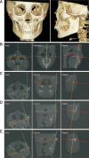
Objective: This study aimed to evaluate the zygomaticotemporal suture (ZTS) maturation, analyze the age distribution patterns of ZTS maturation stages, and investigate the relationship between ZTS and cervical vertebral maturation (CVM).
Methods: A total of 261 patients who underwent cone-beam computed tomography (112 males, mean age, 13.1 ± 3.3 years; 149 females, mean age, 13.7 ± 3.1 years) were examined to evaluate the ZTS stages. The ZTS stages were defined based on a modified method from previous studies on zygomaticomaxillary sutures. Differences between groups and correlations between indicators were analyzed using the Spearman correlation test, intraclass coefficient of correlation (ICC), one-way analysis of variance and rank sum test. Statistical significance was set at p < 0.05. The diagnostic value of CVM stages in identifying ZTS maturation stages was evaluated using positive likelihood ratios (LRs).
Results: A positive relationship was found between the ZTS and CVM stage (r = 0.747, ICC = 0.621, p < 0.01) and between the ZTS stage and chronological age (r = 0.727, ICC = 0.330, p < 0.01). Positive LRs > 10 were found for several cervical stages (CSs), including CS1 and CS2 for the diagnosis of stage B, CS1 to CS3 for the diagnosis of stages B and C, and CS6 for the diagnosis of stages D and E.
Conclusions: The ZTS maturation stage may be more relevant to the CVM stage than to the chronological age. The CVM stages can be good indicators for clinical decisions regarding maxillary protraction, except for CS4 and CS5.
Keywords: Computed tomography; Maxillary protraction; Vertebral maturation; Zygomaticotemporal suture.
Conflict of interest statement
No potential conflict of interest relevant to this article was reported.
Figures


Similar articles
-
Correlation of skeletal development and midpalatal suture maturation.Eur J Med Res. 2024 Sep 16;29(1):461. doi: 10.1186/s40001-024-02058-1. Eur J Med Res. 2024. PMID: 39285501 Free PMC article.
-
Diagnostic performance of skeletal maturity for the assessment of midpalatal suture maturation.Am J Orthod Dentofacial Orthop. 2015 Dec;148(6):1010-6. doi: 10.1016/j.ajodo.2015.06.016. Am J Orthod Dentofacial Orthop. 2015. PMID: 26672707
-
Correlation assessment of cervical vertebrae maturation stage and mid-palatal suture maturation in an Iranian population.J World Fed Orthod. 2020 Sep;9(3):112-116. doi: 10.1016/j.ejwf.2020.05.004. Epub 2020 Jun 28. J World Fed Orthod. 2020. PMID: 32800572
-
Chronological age range estimation of cervical vertebral maturation using Baccetti method: a systematic review and meta-analysis.Eur J Orthod. 2022 Sep 19;44(5):548-555. doi: 10.1093/ejo/cjac009. Eur J Orthod. 2022. PMID: 35258568 Free PMC article.
-
Stability of transversal correction with hybrid maxillary expansion appliance in bone and tegumental piriformis opening in relation to bone age and maturation of the midpalatal suture.J Clin Exp Dent. 2022 May 1;14(5):e439-e445. doi: 10.4317/jced.59575. eCollection 2022 May. J Clin Exp Dent. 2022. PMID: 35582350 Free PMC article. Review.
Cited by
-
Assessment of Zygomaticomaxillary Suture Maturation in Patients With Unilateral Cleft Lip and Palate: Implications for Orthodontic Treatment Planning.Cureus. 2024 Sep 25;16(9):e70146. doi: 10.7759/cureus.70146. eCollection 2024 Sep. Cureus. 2024. PMID: 39463616 Free PMC article.
References
-
- Celikoglu M, Buyukcavus MH. Changes in pharyngeal airway dimensions and hyoid bone position after maxillary protraction with different alternate rapid maxillary expansion and construction protocols: a prospective clinical study. Angle Orthod. 2017;87:519–25. doi: 10.2319/082316-632.1. https://doi.org/10.2319/082316-632.1. - DOI - PMC - PubMed
-
- Willmann JH, Nienkemper M, Tarraf NE, Wilmes B, Drescher D. Early Class III treatment with Hybrid-Hyrax - Facemask in comparison to Hybrid-Hyrax-Mentoplate - skeletal and dental outcomes. Prog Orthod. 2018;19:42. doi: 10.1186/s40510-018-0239-8. https://doi.org/10.1186/s40510-018-0239-8. 2fda100b26f54c179891b36314ca90b6 - DOI - PMC - PubMed
-
- Al Dayeh A, Williams RA, Trojan TM, Claro WI. Deformation of the zygomaticomaxillary and nasofrontal sutures during bone-anchored maxillary protraction and reverse-pull headgear treatments: an ex-vivo study. Am J Orthod Dentofacial Orthop. 2019;156:745–57. doi: 10.1016/j.ajodo.2018.12.019. https://doi.org/10.1016/j.ajodo.2018.12.019. - DOI - PubMed
-
- Vracar TR, Claro W, Vracar ME, 2nd, Jenkins RS, Bland L, Dayeh AA. Sutural deformation during bone-anchored maxillary protraction. J Oral Biol Craniofac Res. 2021;11:447–50. doi: 10.1016/j.jobcr.2021.05.008. https://doi.org/10.1016/j.jobcr.2021.05.008. - DOI - PMC - PubMed
-
- Herring SW. Mechanical influences on suture development and patency. Front Oral Biol. 2008;12:41–56. doi: 10.1159/000115031. https://www.ncbi.nlm.nih.gov/pmc/articles/PMC2826139/ - DOI - PMC - PubMed
LinkOut - more resources
Full Text Sources
Miscellaneous
