. 2023 Sep;22(9):e13914.
doi: 10.1111/acel.13914. Epub 2023 Jun 25.

Deficiency for scavenger receptors Stabilin-1 and Stabilin-2 leads to age-dependent renal and hepatic depositions of fasciclin domain proteins TGFBI and Periostin in mice

Affiliations

Deficiency for scavenger receptors Stabilin-1 and Stabilin-2 leads to age-dependent renal and hepatic depositions of fasciclin domain proteins TGFBI and Periostin in mice

Thomas Leibing et al. Aging Cell. 2023 Sep.

Abstract

Stabilin-1 (Stab1) and Stabilin-2 (Stab2) are two major scavenger receptors of liver sinusoidal endothelial cells that mediate removal of diverse molecules from the plasma. Double-knockout mice (Stab-DKO) develop impaired kidney function and a decreased lifespan, while single Stabilin deficiency or therapeutic inhibition ameliorates atherosclerosis and Stab1-inhibition is subject of clinical trials in immuno-oncology. Although POSTN and TFGBI have recently been described as novel Stabilin ligands, the dynamics and functional implications of these ligands have not been comprehensively studied. Immunofluorescence, Western Blotting and Simple Western™ as well as in situ hybridization (RNAScope™) and qRT-PCR were used to analyze transcription levels and tissue distribution of POSTN and TGFBI in Stab-KO mice. Stab-POSTN-Triple deficient mice were generated to assess kidney and liver fibrosis and function in young and aged mice. TGFBI and POSTN protein accumulated in liver tissue in Stab-DKO mice and age-dependent in glomeruli of Stabilin-deficient mice despite unchanged transcriptional levels. Stab-POSTN-Triple KO mice showed glomerulofibrosis and a reduced lifespan comparable to Stab-DKO mice. However, alterations of the glomerular diameter and vascular density were partially normalized in Stab-POSTN-Triple KO. TGFBI and POSTN are Stabilin-ligands that are deposited in an age-dependent manner in the kidneys and liver due to insufficient scavenging in the liver. Functionally, POSTN might partially contribute to the observed renal phenotype in Stab-DKO mice. This study provides details on downstream effects how Stabilin dysfunction affects organ function on a molecular and functional level.

Keywords: POSTN; TGFBI; aging; fasciclin; lifespan; mouse; scavenger receptors.


Conflict of interest statement

The authors have declared that no conflict of interest exists.

Figures

FIGURE 1
FIGURE 1
POSTN in kidneys and liver from Stabilin‐deficient animals. (a) Representative photomicrographs of kidney tissue (upper lane: Overview, Scale bar = 100 μm; lower lane: Magnified glomeruli, Scale bar = 50 μm.) co‐stained with DAPI (blue), EMCN (green), and POSTN (red). Dashed white line marks glomeruli. (b) Quantification of average POSTN‐positive area in glomeruli (in % of total glomerular area). (c) Quantification of average glomerular diameter (in μm2). (d) Representative photomicrographs of liver tissue co‐stained with DAPI (blue) and POSTN (red). Quantification of average POSTN‐positive area of total photomicrograph area is shown on the right (in % of total photomicrograph area). N ≥ 5 for all experiments. Ns = not significant, *p < 0.05; **p < 0.01; ***p < 0.001.
FIGURE 2
FIGURE 2
Local POSTN transcription is unaltered despite increased protein levels. (a)Left panel: In situ hybridization and Right graph: rt‐PCR of Kidney (upper panel) and Liver (lower panel) in WT and Stab‐DKO animals for POSTN. (b) Positive Control (Dapß) and negative Ctrl (PPIB). Scale bar = 50 μm. N ≥ 3 for all experiments. Ns = not significant, *p < 0.05; **p < 0.01; ***p < 0.001.
FIGURE 3
FIGURE 3
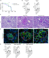
Ablation of POSTN does not influence kidney phenotype in Stab‐DKO mice. (a) Survival curve of Stab‐DKO mice (n = 40) and Stab‐POSTN‐Triple deficient mice (n = 18). (b) Total protein and Albumin levels in different mice strains were assessed (c) Representative photomicrographs of PAS‐stained kidney tissue from different mice strains. (d) Representative photomicrographs of kidney tissue co‐stained with DAPI (blue), EMCN (green), and SPARC (red). Dashed white line marks glomeruli. (e) Quantification of glomerular diameter and EMCN‐positivity in total glomerular area, (f) Quantification of SPARC‐positivity in total glomerular area. Scale bar = 50 μm. n ≥ 5 for all experiments. ns = not significant, *p < 0.05; **p < 0.01; ***p < 0.001.
FIGURE 4
FIGURE 4
Abundance of TGFBI in organs from Stabilin‐deficient animals. (a) Representative photomicrographs of kidney tissue (upper lane: Overview; lower lane: Magnified glomeruli, Scale bar = 50 μm) co‐stained with DAPI (blue), Emcn (green), and TGFBI (magenta). Dashed white line marks glomeruli (b) Quantification of average TGFBI‐positive area in glomeruli (in % of total glomerular area, n ≥ 5). (c) Left panel: In situ hybridization (Scale bar = 50 μm) and right graph: rt‐PCR of kidney tissue in WT and Stab‐DKO animals for TGFBI (n ≥ 3). (d) Representative photomicrographs of liver tissue (upper lane: Overview; lower lane: Magnified pericentral area, Scale bar = 50 μm) co‐stained with DAPI (blue), Emcn (green), and TGFBI (magenta). (e) Quantification of average TGFBI‐positive area of total photomicrograph area (in % of total photomicrograph area, n ≥ 5). (f) Left panel: In situ hybridization (Scale bar = 100 μm) and Right graph: rt‐PCR (n ≥ 3) of kidney tissue in WT and Stab‐DKO animals for TGFBI. ns = not significant, *p < 0.05; **p < 0.01; ***p < 0.001.
FIGURE 5
FIGURE 5
Stab‐TrKO mice share similar TGFBI depositions in liver and kidney with Stab‐DKO mice. (a)Correlation of TGFBI and POSTN abundance from quantified immunofluorescent microphotographs in kidney tissue of DKO (left panels) and WT (right panels) mice. (b) Representative photomicrographs of kidney tissue co‐stained with DAPI (blue), Emcn (green), and TGFBI (magenta). Dashed white line marks glomeruli. Right panel: Quantification of average TGFBI‐positive area in glomeruli (in % of total glomerular area). (c) Representative photomicrographs of liver tissue co‐stained with DAPI (blue), Emcn (green), and TGFBI (magenta). Right panel: Quantification of average TGFBI‐positive area of total photomicrograph area (in % of total photomicrograph area). N ≥ 5 for all experiments. Scale bar = 50 μm. Ns = not significant, *p < 0.05; **p < 0.01; ***p < 0.001.
FIGURE 6
FIGURE 6
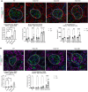
Age‐dependent ligand abundance in glomeruli. (a) Representative photomicrographs of kidney tissue co‐stained with DAPI (blue), Emcn (green), and POSTN (red). Dashed white line marks glomeruli. Lower left panel: Quantification of average POSTN‐positive area in glomeruli (in % of total glomerular area). Lower middle panel: Glomerular POSTN‐positive area in glomeruli (in % of total glomerular area) in 3‐ and 12‐months old mice. Lower right panel: Glomerular area in μm2 in different age groups and genotypes. (b) Representative photomicrographs of kidney tissue co‐stained with DAPI (blue), Emcn (green), and TGFBI (magenta). Dashed white line marks glomeruli. Lower left panel: Quantification of average TGFBI‐positive area in glomeruli (in % of total glomerular area). Lower right panel: Glomerular TGFBI‐positive area in glomeruli (in % of total glomerular area) in 3‐ and 12‐months old mice. n ≥ 5 for all experiments. Scale bar = 50 μm. ns = not significant, *p < 0.05; **p < 0.01; ***p < 0.001.

References

    1. An, J. N. , Kim, H. , Kim, E. N. , Cho, A. , Cho, Y. , Choi, Y. W. , Kim, J. H. , Yang, S. H. , Choi, B. S. , Lim, C. S. , Kim, Y. S. , Kim, K. P. , & Lee, J. P. (2021). Effects of periostin deficiency on kidney aging and lipid metabolism. Aging (Albany NY), 13(19), 22649–22665. 10.18632/aging.203580 - DOI - PMC - PubMed
    1. An, J. N. , Yang, S. H. , Kim, Y. C. , Hwang, J. H. , Park, J. Y. , Kim, D. K. , Kim, J. H. , Kim, D. W. , Hur, D. G. , Oh, Y. K. , Lim, C. S. , Kim, Y. S. , & Lee, J. P. (2019). Periostin induces kidney fibrosis after acute kidney injury via the p38 MAPK pathway. American Journal of Physiology. Renal Physiology, 316(3), F426–f437. 10.1152/ajprenal.00203.2018 - DOI - PubMed
    1. Bhandari, S. , Larsen, A. K. , McCourt, P. , Smedsrød, B. , & Sørensen, K. K. (2021). The scavenger function of liver sinusoidal endothelial cells in health and disease. Frontiers in Physiology, 12. 10.3389/fphys.2021.757469 - DOI - PMC - PubMed
    1. Billings, P. C. , Whitbeck, J. C. , Adams, C. S. , Abrams, W. R. , Cohen, A. J. , Engelsberg, B. N. , Howard, P. S. , & Rosenbloom, J. (2002). The transforming growth factor‐β‐inducible matrix protein βig‐h3 interacts with fibronectin. Journal of Biological Chemistry, 277(31), 28003–28009. 10.1074/jbc.M106837200 - DOI - PubMed
    1. Guerrot, D. , Dussaule, J. C. , Mael‐Ainin, M. , Xu‐Dubois, Y. C. , Rondeau, E. , Chatziantoniou, C. , & Placier, S. (2012). Identification of periostin as a critical marker of progression/reversal of hypertensive nephropathy. PLoS One, 7(3), e31974. 10.1371/journal.pone.0031974 - DOI - PMC - PubMed

Publication types

LinkOut - more resources