Case Reports
. 2023 Jun;29(6):e13380.
doi: 10.1111/srt.13380.

Dermoscopy and high-frequency ultrasound provide diagnostic clues in a gastric adenocarcinoma with cutaneous metastasis as the initial presentation: A case report

Affiliations
Case Reports

Dermoscopy and high-frequency ultrasound provide diagnostic clues in a gastric adenocarcinoma with cutaneous metastasis as the initial presentation: A case report

Juncheng Wang et al. Skin Res Technol. 2023 Jun.
No abstract available


Conflict of interest statement

The authors declare they have no conflict of interest.

Figures
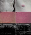
FIGURE 1
FIGURE 1
(A and B) The patient presented with multiple flesh‐colored and erythematous skin nodules on his abdomen, left chest and right waist. (C and D) Dermoscopic examination revealed dilated linear irregular and branching vessels with a reticular distribution on a red background. (E and F) Ultrasonographic examination using 50 and 20 MHz probes demonstrated multiple longitudinal hypoechoic areas in the superficial dermis, connected to larger hypoechoic areas in the deep dermis. These areas exhibited an irregular shape, an ill‐defined border, and infiltration into the subcutaneous tissue.
FIGURE 2
FIGURE 2
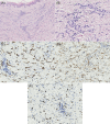
(A) Histopathological examination revealed scattered infiltration of atypical cells among the collagen fibers in the dermis (HE, ×100). (B) The atypical cells were pleomorphic and hyperchromatic (HE, ×400). (C and D) Immunohistochemical staining showed positive expression of CK and MPO in the atypical cells (×400). (E) The Ki‐67 index was determined to be 80% (×400).
FIGURE 3
FIGURE 3
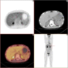
A whole‐body fluorodeoxyglucose positron emission tomography/computed tomography scan revealed thickening of the gastric wall in the cardia region, accompanied by increased metabolic activity (SUVmax 3.6).

Similar articles

Cited by

  • High-Frequency and Ultra-High-Frequency Ultrasound in Dermatologic Diseases and Aesthetic Medicine.
    Argalia G, Reginelli A, Molinelli E, Russo A, Michelucci A, Sechi A, Marzano AV, Desyatnikova S, Fogante M, Patanè V, Granieri G, Tagliati C, Rizzetto G, De Simoni E, Matteucci M, Candelora M, Lanza C, Ventura C, Carboni N, Esposito R, Esposito S, Paolinelli M, Esposto E, Lanni G, Lucidi Pressanti G, Giorgi C, Principi F, Rebonato A, Malinowska SP, Mlosek RK, Giuseppetti GM, Dini V, Romanelli M, Offidani A, Cappabianca S, Wortsman X, Simonetti O. Argalia G, et al. Medicina (Kaunas). 2025 Jan 26;61(2):220. doi: 10.3390/medicina61020220. Medicina (Kaunas). 2025. PMID: 40005337 Free PMC article. Review.

References

    1. Hu SCS, Ke CLK, Chen GS, Cheng ST. Dermoscopic vascular network in cutaneous metastases from nasopharyngeal carcinoma. J Eur Acad Dermatol Venereol. 2016;30:e29‐e31. - PubMed
    1. Kamińska‐Winciorek G, Pilśniak A, Piskorski W, Wydmański J. Skin metastases in the clinical and dermoscopic aspects. Semin Oncol. 2022;49:160‐169. - PubMed
    1. Ghosh J, Arun I, Ganguly A, Ganguly S. Cutaneous metastases in a patient with adenocarcinoma of the stomach. Indian J Dermatol, Venereol Leprol. 2021;87:699‐701. - PubMed
    1. Chernoff KA, Marghoob AA, Lacouture ME, Deng L, Busam KJ, Myskowski PL. Dermoscopic findings in cutaneous metastases. JAMA Dermatol. 2014;150:429‐433. - PubMed
    1. Ertop Doğan P, Akay BN, Hazinedar E, Koca R. Clinical and dermoscopic findings of cutaneous metastases: a case series. Int J Dermatol. 2022;61:e351‐e3. - PubMed

MeSH terms