. 2023 Aug 17;11(4):e0521122.
doi: 10.1128/spectrum.05211-22. Epub 2023 Jun 26.

FAM46C Is an Interferon-Stimulated Gene That Inhibits Lentiviral Particle Production by Modulating Autophagy

Affiliations

FAM46C Is an Interferon-Stimulated Gene That Inhibits Lentiviral Particle Production by Modulating Autophagy

Marilena Mancino et al. Microbiol Spectr. .

Abstract

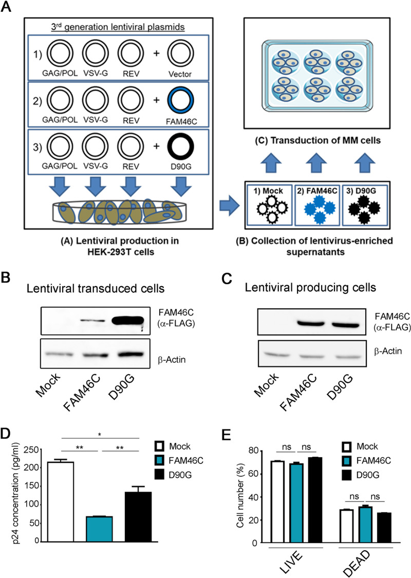
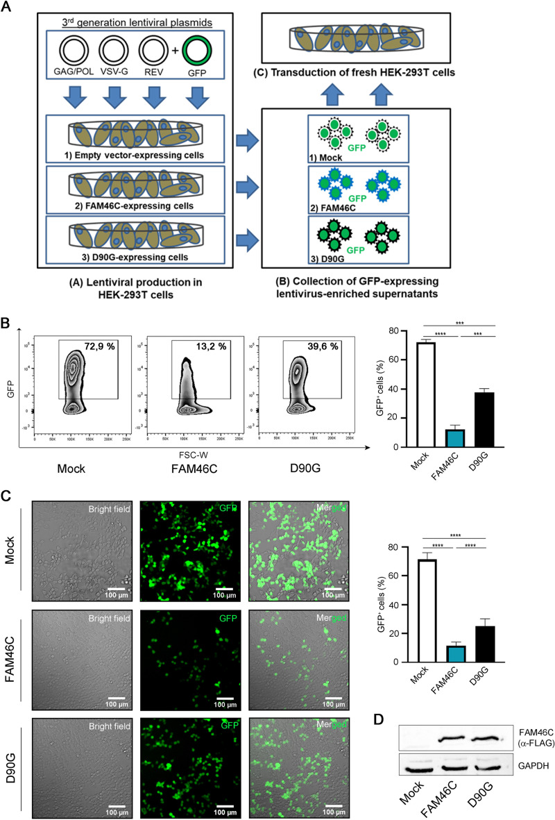
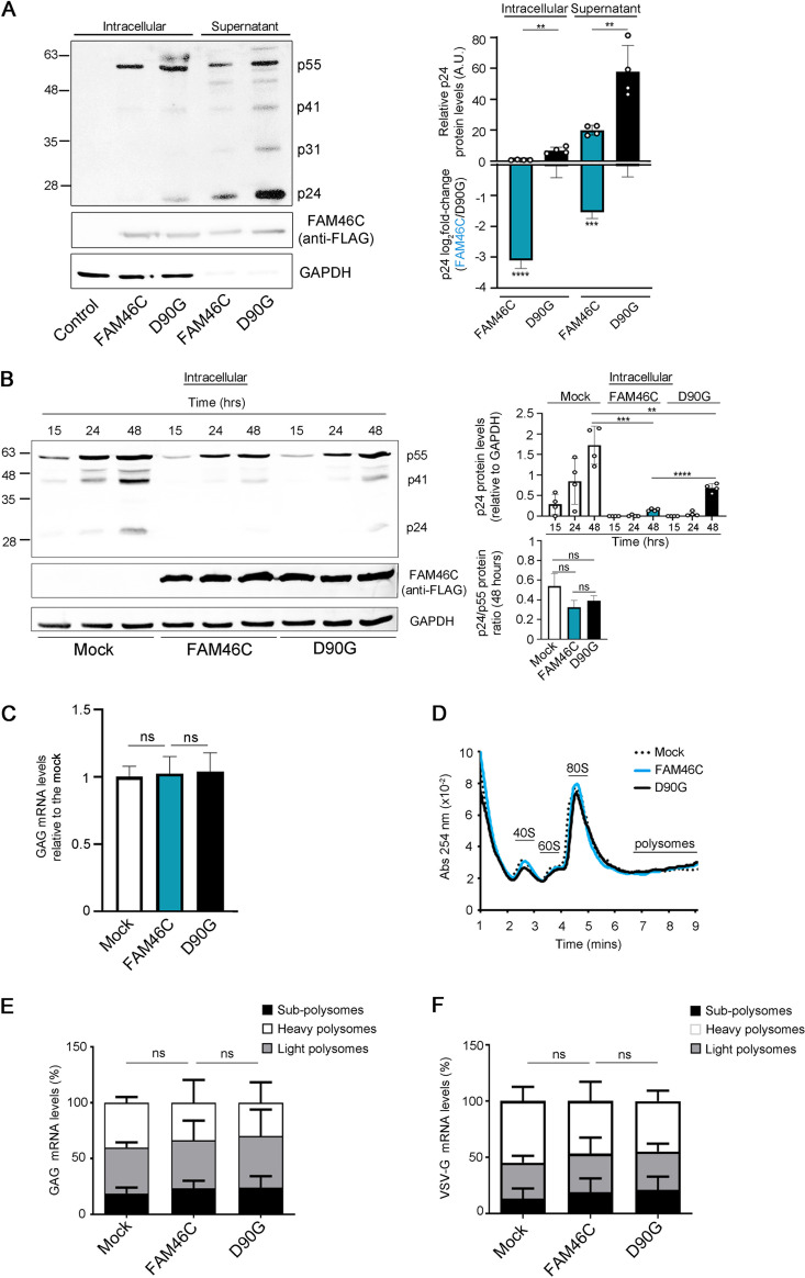
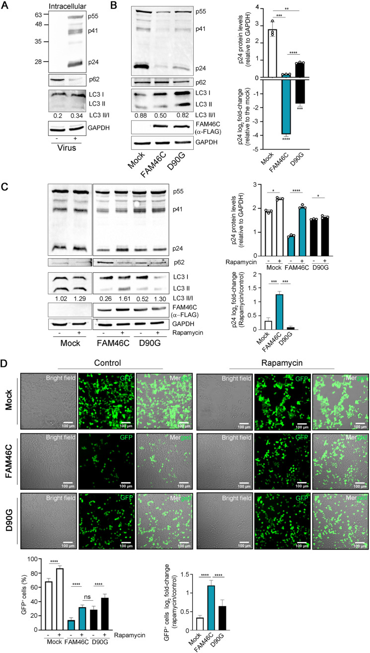
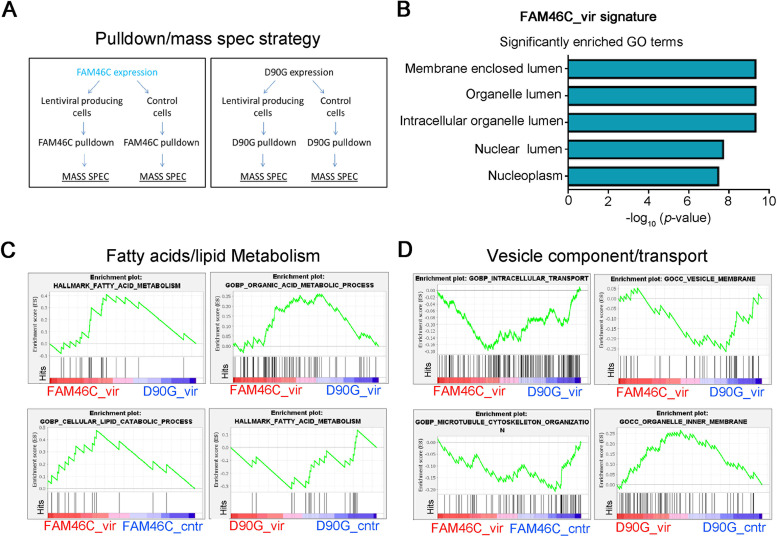
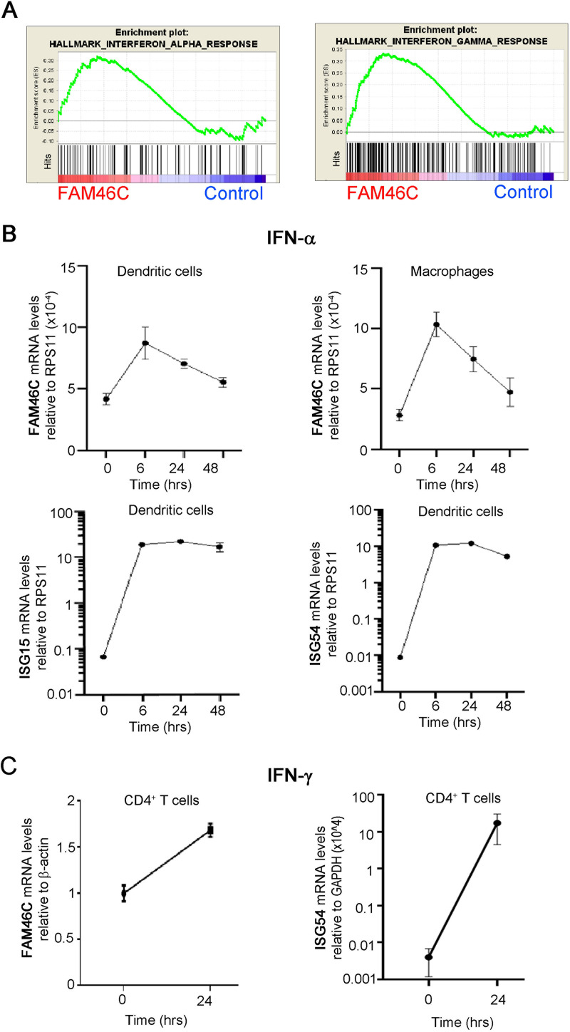
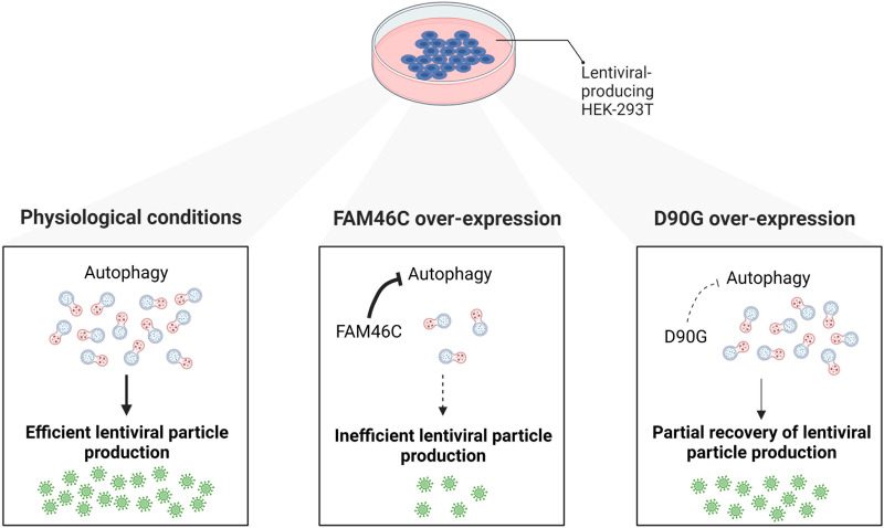
FAM46C is a multiple myeloma (MM) tumor suppressor whose function is only starting to be elucidated. We recently showed that in MM cells FAM46C triggers apoptosis by inhibiting autophagy and altering intracellular trafficking and protein secretion. To date, both a physiological characterization of FAM46C role and an assessment of FAM46C-induced phenotypes outside of MM are lacking. Preliminary reports suggested an involvement of FAM46C with regulation of viral replication, but this was never confirmed. Here, we show that FAM46C is an interferon-stimulated gene and that the expression of wild-type FAM46C in HEK-293T cells, but not of its most frequently found mutant variants, inhibits the production of both HIV-1-derived and HIV-1 lentiviruses. We demonstrate that this effect does not require transcriptional regulation and does not depend on inhibition of either global or virus-specific translation but rather mostly relies on FAM46C-induced deregulation of autophagy, a pathway that we show to be required for efficient lentiviral particle production. These studies not only provide new insights on the physiological role of the FAM46C protein but also could help in implementing more efficient antiviral strategies on one side and lentiviral particle production approaches on the other. IMPORTANCE FAM46C role has been thoroughly investigated in MM, but studies characterizing its role outside of the tumoral environment are still lacking. Despite the success of antiretroviral therapy in suppressing HIV load to undetectable levels, there is currently no HIV cure, and treatment is lifelong. Indeed, HIV continues to be a major global public health issue. Here, we show that FAM46C expression in HEK-293T cells inhibits the production of both HIV and HIV-derived lentiviruses. We also demonstrate that such inhibitory effect relies, at least in part, on the well-established regulatory role that FAM46C exerts on autophagy. Deciphering the molecular mechanism underlying this regulation will not only facilitate the understanding of FAM46C physiological role but also give new insights on the interplay between HIV and the cellular environment.

Keywords: FAM46C; HIV-1; TENT5C; autophagy; interferons; lentiviruses; viral production.


Conflict of interest statement

The authors declare no conflict of interest.

Figures

FIG 1
FIG 1
FAM46C inhibits lentiviral particle production in cis. (A) Cartoon depicting our viral particle production and transduction approach. (Subpanel A) HEK-293T cells were transfected with plasmids encoding for the lentiviral GAG/POL, VSV-G, and REV genes, together with plasmids encoding for either the FAM46C gene, the D90G mutant allele, or an empty vector. (Subpanels B and C) The viral particles produced were then collected from the supernatant (subpanel B) and used to infect OPM2 MM cells (subpanel C). (B) Western blot analysis showing FAM46C and D90G protein levels in OPM2 MM cells. OPM2 cells were transduced with FAM46C-, D90G-, or empty vector-expressing lentiviruses as described in panel A. Cytoplasmic lysates derived from the samples were subjected to SDS-PAGE and Western blotting with α-FLAG antibodies. β-Actin was used as a loading control. (C) Western blot analysis showing FAM46C and D90G protein levels in virus-producing HEK-293T cells. HEK-293T cells were transfected to produce lentiviral vectors expressing either FAM46C, the D90G mutant allele, or an empty vector, as described in panel A. Samples were then prepared as described in panel B. β-Actin was used as a loading control. (D) p24 concentrations in the lentivirus-enriched supernatants produced by HEK-293T cells. The lentivirus-containing supernatants produced by the cells described in panel C were analyzed for p24 levels by ELISA. (E) Percentage of live and dead HEK-293T cells during lentiviral production. The cells described in panel C were used to assess cell viability through flow cytometry. Blots are representative of three independent experiments. Histograms represent the means ± the SD of three independent experiments. Statistical P values were calculated using double-tailed unpaired t tests. ns, P > 0.05; **, P < 0.01.
FIG 2
FIG 2
FAM46C inhibits lentiviral particle production in trans. (A) Cartoon depicting our viral particle production and transduction approach. (Subpanel A) HEK-293T cells expressing either an empty vector, FAM46C, or the D90G mutant were transfected with plasmids encoding for the lentiviral GAG/POL, VSV-G, and REV genes and GFP. (Subpanels B and C) The GFP-expressing lentiviral particles produced were then collected from the supernatant (subpanel B) and used to infect fresh HEK-293T cells (subpanel C). (B) Quantitation of GFP+ HEK-293T cells after transduction with GFP-expressing lentiviruses. GFP-expressing lentiviruses produced in FAM46C-, D90G-, or empty vector-expressing HEK-293T cells were used to infect fresh HEK-293T cells as described in panel A. The number of GFP+ cells was then assessed by flow cytometry. (Left) Representative zebra plots showing the percentage of GFP+ cells. (Right) Histograms representing the means ± the SD of three independent experiments. (C) Fluorescence microscopy analysis of HEK-293T cells transduced with GFP-expressing lentiviruses. The cells in panel B were analyzed by fluorescence microscopy. (Left) Representative confocal microscopy images. (Right) Histograms representing the mean percentage of GFP+ cells ± the SD of 12 independent fields of view. (D) Western blot analysis showing FAM46C and D90G protein levels in virus-producing HEK-293T cells. HEK-293T cells were transfected to produce GFP-expressing lentiviral particles in the presence of either FAM46C, the D90G mutant, or an empty vector, as described in panel A. Cytoplasmic lysates derived from the samples were subjected to SDS-PAGE and Western blotting with α-FLAG-antibodies. GAPDH was used as a loading control. Statistical P values were calculated using double-tailed unpaired t tests. ***, P < 0.001; ****, P < 0.0001.
FIG 3
FIG 3
FAM46C inhibits lentiviral particle production by regulating viral gene expression at the posttranslational level. (A, left) Representative western blot showing the levels of viral proteins both in the intracellular compartment and in the supernatant of viral-producing HEK-293T cells. HEK-293T cells were transfected as described in Fig. 1A. Protein samples were extracted and subjected to SDS-PAGE and western blotting with the antiserum of an HIV-1-infected patient. GAPDH was used as a loading control. (Right) Histograms representing absolute and relative p24 levels. (B, left) Representative western blot showing the levels of intracellular GAG proteins in virus-producing HEK-293T cells. HEK-293T cells were transfected as described in Fig. 2A, and GAG protein levels were detected by western blotting following SDS-PAGE on protein lysates at the indicated time points after transfection using α-p24 antibodies. GAPDH was used as a loading control. (Right) Histograms representing relative p24 levels and p24/p55 ratios. (C) GAG mRNA levels in HEK-293T lentivirus-producing cells. HEK-293T cells were transfected as described in Fig. 2A, and GAG mRNA levels were determined by RT-qPCR. (D) Polysome profiles of HEK-293T lentivirus-producing cells. The cells in panel C were lysed, and polysome profiles were isolated and analyzed. (E and F) Levels of GAG (E) and VSV-G (F) viral transcripts in different polysome fractions of lentivirus-producing HEK-293T cells. Polysome fractions from the samples described in panel D were purified and assessed for transcript abundance. Histograms represent the means ± the SD of three independent experiments. Blots are representative of three independent experiments. Statistical P values were calculated using double-tailed unpaired t tests. ns, P > 0.05.
FIG 4
FIG 4
Autophagy is required for FAM46C-induced inhibition of viral particle production. (A) Representative western blot showing the levels autophagic markers and Gag proteins in virus-producing HEK-293T cells. HEK-293T cells were transfected with lentiviral envelope and packaging plasmids only. Cytoplasmic lysates derived from the samples were subjected to SDS-PAGE and western blotting with α-p24 and α-p62 antibodies. GAPDH was used as loading control. (B, left) Representative western blot showing the levels autophagic markers and Gag proteins in virus-producing HEK-293T cells. HEK-293T cells were transfected as described in Fig. 2B. Cytoplasmic lysates derived from the samples were subjected to SDS-PAGE and western blotting with α-p62 and α-LC3-II α-p24 and α-FLAG antibodies. GAPDH was used as a loading control. (Right) Histograms representing absolute and relative p24 levels. (C, left) Representative western blot showing the levels autophagic markers and Gag proteins in HEK-293T cells transfected as in panel A but either treated or not treated 24 h posttransfection with 100 nM rapamycin for an additional 24 h. Samples were prepared as in panel A. (Right) Histograms representing absolute and relative p24 levels. (D) Fluorescence microscopy analysis of HEK-293T cells transduced with GFP-expressing lentiviruses. GFP-expressing lentiviruses produced in FAM46C-, D90G-, or empty vector-expressing HEK-293T cells either treated or not treated with rapamycin were used to infect fresh HEK-293T cells. (Top) Representative confocal microscopy images. (Bottom) Histograms representing either the mean percentage or the log2-fold change of GFP+ cells ± the SD of 14 independent fields of view. Statistical P values were calculated using double-tailed unpaired t tests. ns, P > 0.05; ***, P < 0.001; ****, P < 0.0001.
FIG 5
FIG 5
During lentiviral production, the interactome of FAM46C is enriched in proteins involved in organelle homeostasis and intracellular trafficking. (A) Cartoon depicting our pulldown/mass spectrometry approach. HEK-293T cells were transfected with plasmids encoding FAM46C or the D90G mutant either in the presence or in the absence of lentiviral packaging and envelope plasmids. FAM46C and D90G proteins were then coimmunoprecipitated, and their interactomes were analyzed by mass spectrometry. (B) GO terms significantly enriched for proteins that interact more with FAM46C during viral production. (C and D) GSEA on the interactomes of FAM46C and D90G during lentiviral production. By using GSEA, we compared the interactome of FAM46C with that of the D90G mutant during viral particle production (FAM46C_vir/D90G_vir) (top) and also each of the two interactomes with their counterpart in the absence of viral particle production (FAM46C_vir/FAM46C_cntr and D90G_vir/D90G_cntr) (bottom), highlighting fatty acid/lipid metabolism (C) and in vesicle component/transport (D) signatures.
FIG 6
FIG 6
FAM46C is stimulated by both type I and type II IFNs. (A) GSEA on microarray data from OPM2 MM cells expressing either FAM46C or a mock control. Signatures related to IFN responses are shown. (B) FAM46C expression levels upon IFN-α administration. Dendritic cells and macrophages were isolated from the peripheral blood of healthy donors and stimulated with IFN-α for 48 h. At the time points indicated, the cells were harvested, and RNA was extracted. Transcript levels were determined by RT-qPCR with normalization on RPS11. ISG15 and ISG54 transcript levels were used as controls. (C) FAM46C expression levels upon IFN-γ administration. CD4+ T cells were isolated as described for panel B and stimulated for 24 h with IFN-γ. Transcript levels were determined by RT-qPCR with normalization on β-actin. ISG54 transcript levels were used as a control. Graphs represent the means ± the SD of three independent experiments.
FIG 7
FIG 7
Cartoon depicting our working model. FAM46C expression inhibits lentiviral particle production by negatively regulating autophagy. (Left) In HEK-293T cells, the physiological levels of autophagy favor lentiviral replication and particle production. (Center) When FAM46C is overexpressed autophagy is dampened and virus replication and particle production are consequently reduced. (Right) The expression of the D90G mutant allele partially suppresses FAM46C-induced autophagy-dependent inhibition of viral replication and particle production.

Similar articles

Cited by

References

    1. Bolli N, Avet-Loiseau H, Wedge DC, Van Loo P, Alexandrov LB, Martincorena I, Dawson KJ, Iorio F, Nik-Zainal S, Bignell GR, Hinton JW, Li Y, Tubio JMC, McLaren S, O’ Meara S, Butler AP, Teague JW, Mudie L, Anderson E, Rashid N, Tai Y-T, Shammas MA, Sperling AS, Fulciniti M, Richardson PG, Parmigiani G, Magrangeas F, Minvielle S, Moreau P, Attal M, Facon T, Futreal PA, Anderson KC, Campbell PJ, Munshi NC. 2014. Heterogeneity of genomic evolution and mutational profiles in multiple myeloma. Nat Commun 5:2997. doi:10.1038/ncomms3997. - DOI - PMC - PubMed
    1. Lohr JG, Stojanov P, Carter SL, Cruz-Gordillo P, Lawrence MS, Auclair D, Sougnez C, Knoechel B, Gould J, Saksena G, Cibulskis K, McKenna A, Chapman MA, Straussman R, Levy J, Perkins LM, Keats JJ, Schumacher SE, Rosenberg M, Getz G, Golub TR, Anderson KC, Richardson P, Krishnan A, Lonial S, Kaufman J, Siegel DS, Vesole DH, Roy V, Rivera CE, Rajkumar SV, Kumar S, Fonseca R, Ahmann GJ, Bergsagel PL, Stewart AK, Hofmeister CC, Efebera YA, Jagannath S, Chari A, Trudel S, Reece D, Wolf J, Martin T, Zimmerman T, Rosenbaum C, Jakubowiak AJ, Lebovic D, Vij R, Stockerl-Goldstein K, Multiple Myeloma Research Consortium . 2014. Widespread genetic heterogeneity in multiple myeloma: implications for targeted therapy. Cancer Cell 25:91–101. doi:10.1016/j.ccr.2013.12.015. - DOI - PMC - PubMed
    1. Chapman MA, Lawrence MS, Keats JJ, Cibulskis K, Sougnez C, Schinzel AC, Harview CL, Brunet J-P, Ahmann GJ, Adli M, Anderson KC, Ardlie KG, Auclair D, Baker A, Bergsagel PL, Bernstein BE, Drier Y, Fonseca R, Gabriel SB, Hofmeister CC, Jagannath S, Jakubowiak AJ, Krishnan A, Levy J, Liefeld T, Lonial S, Mahan S, Mfuko B, Monti S, Perkins LM, Onofrio R, Pugh TJ, Rajkumar SV, Ramos AH, Siegel DS, Sivachenko A, Stewart AK, Trudel S, Vij R, Voet D, Winckler W, Zimmerman T, Carpten J, Trent J, Hahn WC, Garraway LA, Meyerson M, Lander ES, Getz G, Golub TR. 2011. Initial genome sequencing and analysis of multiple myeloma. Nature 471:467–472. doi:10.1038/nature09837. - DOI - PMC - PubMed
    1. Barbieri M, Manzoni M, Fabris S, Ciceri G, Todoerti K, Simeon V, Musto P, Cortelezzi A, Baldini L, Neri A, Lionetti M. 2016. Compendium of FAM46C gene mutations in plasma cell dyscrasias. Br J Haematol 174:642–645. doi:10.1111/bjh.13793. - DOI - PubMed
    1. Boyd KD, Ross FM, Walker BA, Wardell CP, Tapper WJ, Chiecchio L, Dagrada G, Konn ZJ, Gregory WM, Jackson GH, Child JA, Davies FE, Morgan GJ, NCRI Haematology Oncology Studies Group . 2011. Mapping of chromosome 1p deletions in myeloma identifies FAM46C at 1p12 and CDKN2C at 1p32.3 as being genes in regions associated with adverse survival. Clin Cancer Res 17:7776–7784. doi:10.1158/1078-0432.CCR-11-1791. - DOI - PMC - PubMed

Publication types

LinkOut - more resources