. 2023 Aug 17;11(4):e0278022.
doi: 10.1128/spectrum.02780-22. Epub 2023 Jun 26.

Population Dynamics of Intestinal Enterococcus Modulate Galleria mellonella Metamorphosis

Affiliations

Population Dynamics of Intestinal Enterococcus Modulate Galleria mellonella Metamorphosis

Hyun Gi Kong et al. Microbiol Spectr. .

Abstract

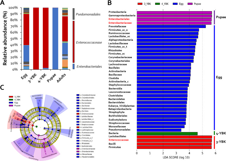
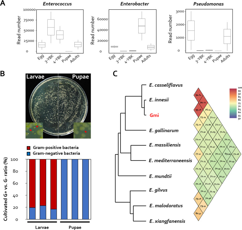
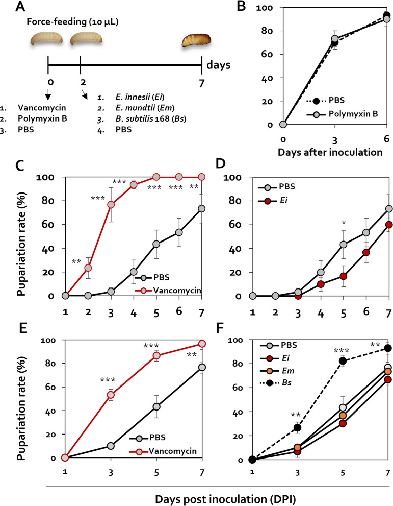
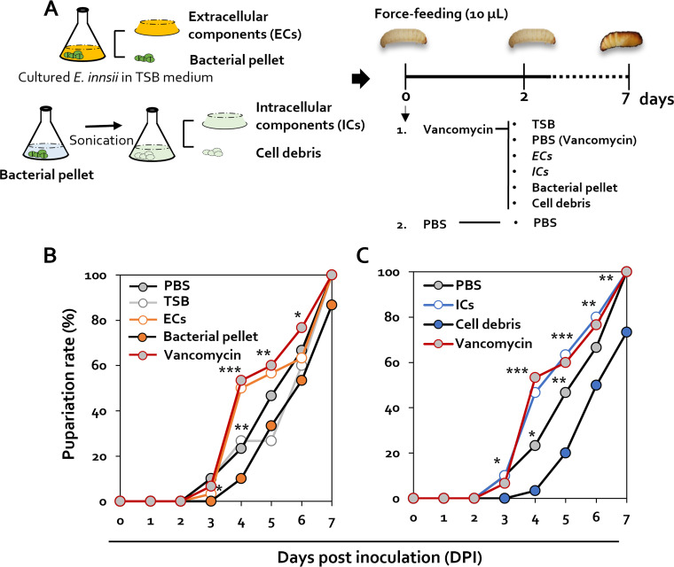
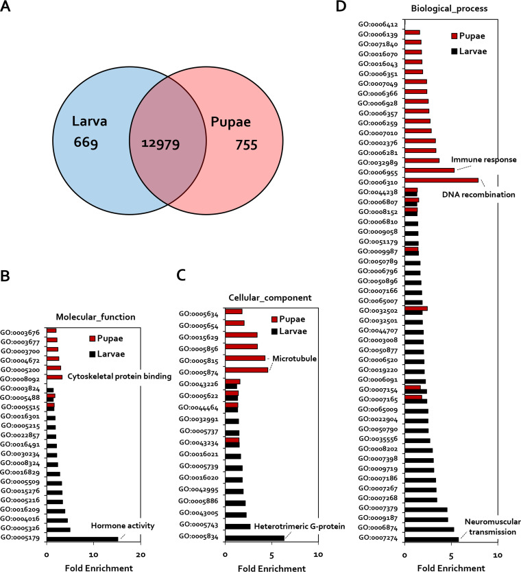
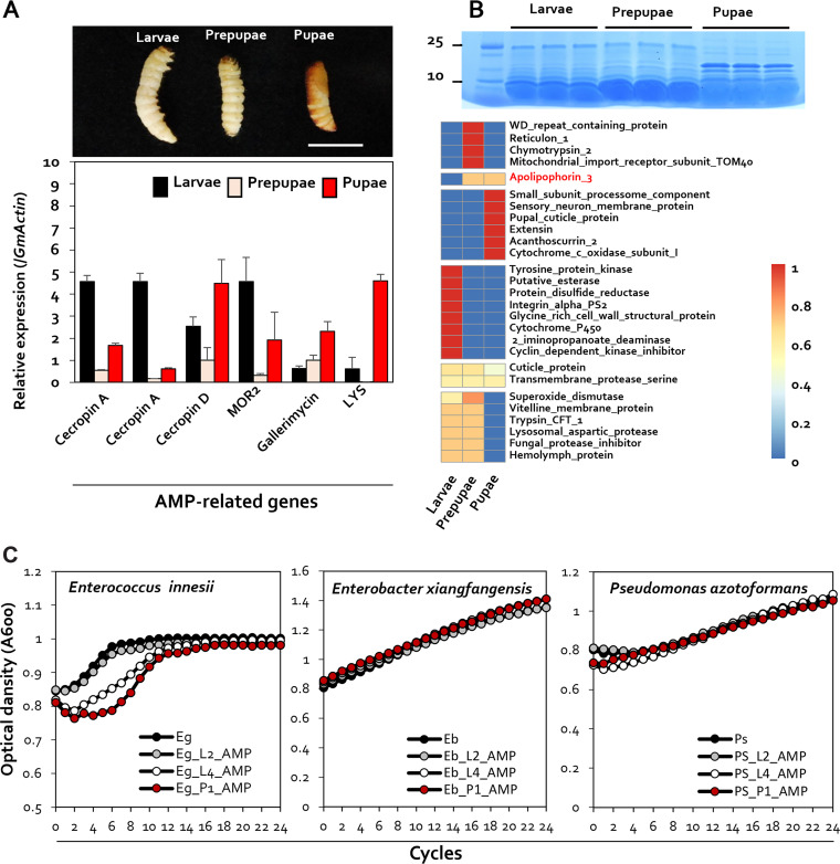
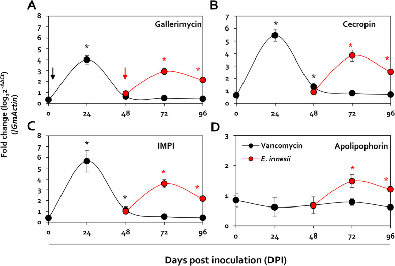
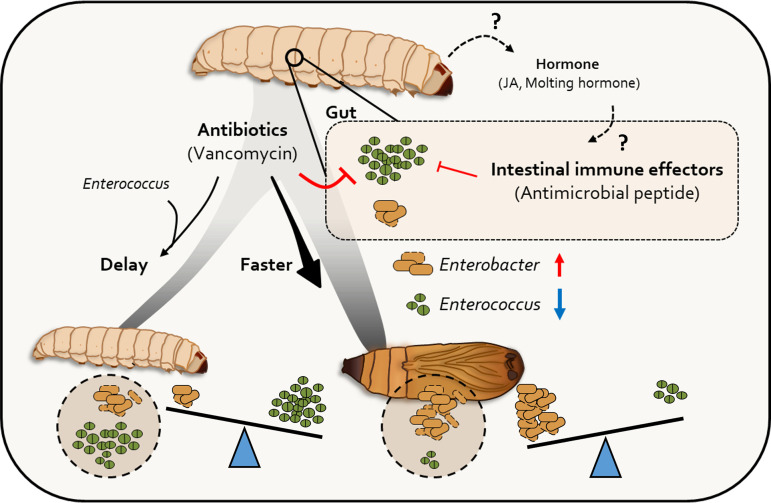
Microbes found in the digestive tracts of insects are known to play an important role in their host's behavior. Although Lepidoptera is one of the most varied insect orders, the link between microbial symbiosis and host development is still poorly understood. In particular, little is known about the role of gut bacteria in metamorphosis. Here, we explored gut microbial biodiversity throughout the life cycle of Galleria mellonella, using amplicon pyrosequencing with the V1 to V3 regions, and found that Enterococcus spp. were abundant in larvae, while Enterobacter spp. were predominant in pupae. Interestingly, eradication of Enterococcus spp. from the digestive system accelerated the larval-to-pupal transition. Furthermore, host transcriptome analysis demonstrated that immune response genes were upregulated in pupae, whereas hormone genes were upregulated in larvae. In particular, regulation of antimicrobial peptide production in the host gut correlated with developmental stage. Certain antimicrobial peptides inhibited the growth of Enterococcus innesii, a dominant bacterial species in the gut of G. mellonella larvae. Our study highlights the importance of gut microbiota dynamics on metamorphosis as a consequence of the active secretion of antimicrobial peptides in the G. mellonella gut. IMPORTANCE First, we demonstrated that the presence of Enterococcus spp. is a driving force for insect metamorphosis. RNA sequencing and peptide production subsequently revealed that antimicrobial peptides targeted against microorganisms in the gut of Galleria mellonella (wax moth) did not kill Enterobacteria species, but did kill Enterococcus species, when the moth was at a certain stage of growth, and this promoted moth pupation.

Keywords: host-microbe interactions; insect metamorphosis; microbiota alteration.


Conflict of interest statement

The authors declare no conflict of interest.

Figures

FIG 1
FIG 1
Changes in symbiont diversity across life stages of G. mellonella. (A) Relative abundance (percentage) of bacterial genera at each developmental stage (n = 8). (B) A histogram of the linear discriminant analysis scores reveals the most differentially abundant taxa among different development stages: green represents 3rd instar larvae, red represents 4th instar larvae, blue represents eggs, and purple represents pupa-enriched taxa. (C) Cladogram generated by the LEfSe method indicating the phylogenetic distribution of gut microbiota in G. mellonella across the development stages from 500 exploratory bootstrap trials. Bar, 0.01 changes per site. The 3rd instar larvae are marked with 3-YBK and 4th instar larvae with 3-YBK.
FIG 2
FIG 2
Isolation of symbionts from G. mellonella larvae and pupae. (A) Read number of OTU corresponding to Enterococcus, Enterobacter, and Providencia at each developmental stage (n = 12). (B) Investigation of the distribution of bacteria cultured from G. mellonella larvae and pupae. Representative bacterial colonies obtained after plating on TSA medium are shown. (C) The average nucleotide identity (ANI) value of the entire genome sequence of the genus Enterococcus sp. and the whole-genome information of the isolate were determined using the NCBI database (https://www.ncbi.nlm.nih.gov/).
FIG 3
FIG 3
Changes in metamorphosis in the presence of Enterococcus innesii. (A) Schematic diagram showing the regimen for treatment of Galleria mellonella larvae with antibiotics and E. innesii. (B) Pupation changes after polymyxin B (100 μg/mL) treatments were confirmed using 4th instar larvae. Pupation rates of larva challenged with PBS (black circles on black dotted line) or polymyxin B (gray circles on black line) are shown. (C) The pupal changes of E. innesii (1 × 105 CFU/larva) treatment and vancomycin (100 μg/mL) treatment were confirmed using 4th instar larvae. Antibiotics and bacteria were fed to the larvae through the mouth, and the degree of pupation was measured by culturing at 30°C for 8 days after feeding. To analyze the recovery of metamorphosis caused by E. innesii after antibiotic treatment, a bacterial suspension was fed to larvae 2 days after the antibiotic feeding. Treatment groups were treated with vancomycin (gray circles on black line), control (gray circles on black line), and E. innesii treatment after vancomycin treatment (gray circles on red line). (D) Analysis of changes in pupation by E. innesii treatment after vancomycin treatment. Shown are results from treatment with E. innesii (e.g., black circles on black line) and the control (gray circles on black line). (E and F) Analysis of changes in pupation by E. innesii (Ei), E. mundtii (Em), and Bacillus subtilis (Bs) treatment. All experiments were performed in triplicate with 30 larvae per replicate.
FIG 4
FIG 4
Pupation changes associated with specific Enterococcus cell components. (A) Schematic diagram showing the methods used for isolating the components of E. innesii used in the treatment of G. mellonella larvae. (B) Pupation changes after treatments with extracellular components (ECs), TSB, PBS, bacterial pellet, and vancomycin were confirmed using 4th instar larvae. (C) Pupation changes after treatments with intracellular components (ICs), PBS, cell debris, and vancomycin were confirmed using 4th instar larvae. All experiments were performed in triplicate with 10 larvae per replicate.
FIG 5
FIG 5
Profiling of differentially expressed genes and Gene Ontology clustering in the G. mellonella following developmental stage. (A) Venn diagram showing differentially expressed genes with the smallest false-discovery rate (FDR): >2. (B to D) Gene Ontology term classification of genes upregulated in larvae or pupae related to molecular function (B), cellular components (C), and biological process (D). The hormone activity (GO:0005179) and immune response (GO:0006955) were significant changes of the transcriptomic pattern between larvae and pupae. Enriched transcripts of pupae are shown in red. Enriched transcripts of larvae are shown in black.
FIG 6
FIG 6
Expression of G. mellonella antimicrobial peptides at different developmental stages. (A) Quantification of gene expression of the antimicrobial peptide genes coding for cecropin A, cecropin D, MOR2, gallerimycin, and LYS in larvae, prepupae, and pupae. The housekeeping gene coding for G. mellonella actin (GmActin) was used for normalization. Error bars represent the mean ± standard error of the mean (SEM). Sample size: n = 5 G. mellonella larvae per treatment. (B) SDS-PAGE analysis of peptide patterns for proteins of 25 kDa or less in the larval, prepupal, and pupal stages of G. mellonella. Shown is a heat map of differentially expressed proteins from larvae, prepupae, and pupae. Proteomic analysis was performed via LC-ESI-Q-TOF, and final qualitative analysis was performed via MS/MS analysis and UniProt data. (C) Antimicrobial activity of 2nd instar larval, 4th instar larval, and pupal antimicrobial peptide extracts against Enterococcus innesii, Enterobacter xianfgangensis, and Pseudomonas azotoformans. The figure shows bacterial growth curves in the presence of antimicrobial peptides extracted from G. mellonella at different developmental stages.
FIG 7
FIG 7
Analysis of gene expression changes in larvae, following vancomycin treatment, through real-time quantitative PCR (RT-qPCR). Shown are levels of expression of the IMPI, cecropin D, gallerimycin, and apolipophorin III genes, which increase in larvae after being fed with vancomycin, shown as fold change (log2 threshold cycle [2−ΔΔCT]) by RT-qPCR. The gene coding for β-actin was used as the reference gene. E. innesii was treated 48 h after antibiotic treatment, and antibiotic treatment and E. innesii treatment are indicated by black and red arrows, respectively.
FIG 8
FIG 8
Role of gut microbiota during metamorphosis. The transition of gut microbiota accelerates the pupation rate. Antimicrobial peptide production, which influences gut microbiota transition, is programmed as a biological clock at the early pupation stage. Antimicrobial peptides reduce the level of Enterococcus, resulting in the induction of G. mellonella pupation.

References

    1. Koch H, Schmid-Hempel P. 2011. Socially transmitted gut microbiota protect bumble bees against an intestinal parasite. Proc Natl Acad Sci USA 108:19288–19292. doi: 10.1073/pnas.1110474108. - DOI - PMC - PubMed
    1. Chen B, Teh BS, Sun C, Hu S, Lu X, Boland W, Shao Y. 2016. Biodiversity and activity of the gut microbiota across the life history of the insect herbivore Spodoptera littoralis. Sci Rep 6:29505. doi: 10.1038/srep29505. - DOI - PMC - PubMed
    1. Fisher RM, Henry LM, Cornwallis CK, Kiers ET, West SA. 2017. The evolution of host-symbiont dependence. Nat Commun 8:15973. doi: 10.1038/ncomms15973. - DOI - PMC - PubMed
    1. Selek G, Ebru G, Parlak O. 2014. Programmed cell death in the digestive canal of Bombyx mori (Lepidoptera: Bombycidae) during prepupal period. IUFS J Biol 73:17–29.
    1. Lemaitre B, Miguel-Aliaga I. 2013. The digestive tract of Drosophila melanogaster. Annu Rev Genet 47:377–404. doi: 10.1146/annurev-genet-111212-133343. - DOI - PubMed

Publication types

Substances

LinkOut - more resources