. 2023 Jul 27;97(7):e0196422.
doi: 10.1128/jvi.01964-22. Epub 2023 Jun 26.

Reverse Genetic Assessment of the Roles Played by the Spike Protein and ORF3 in Porcine Epidemic Diarrhea Virus Pathogenicity

Affiliations

Reverse Genetic Assessment of the Roles Played by the Spike Protein and ORF3 in Porcine Epidemic Diarrhea Virus Pathogenicity

Claudia Kristen-Burmann et al. J Virol. .

Abstract

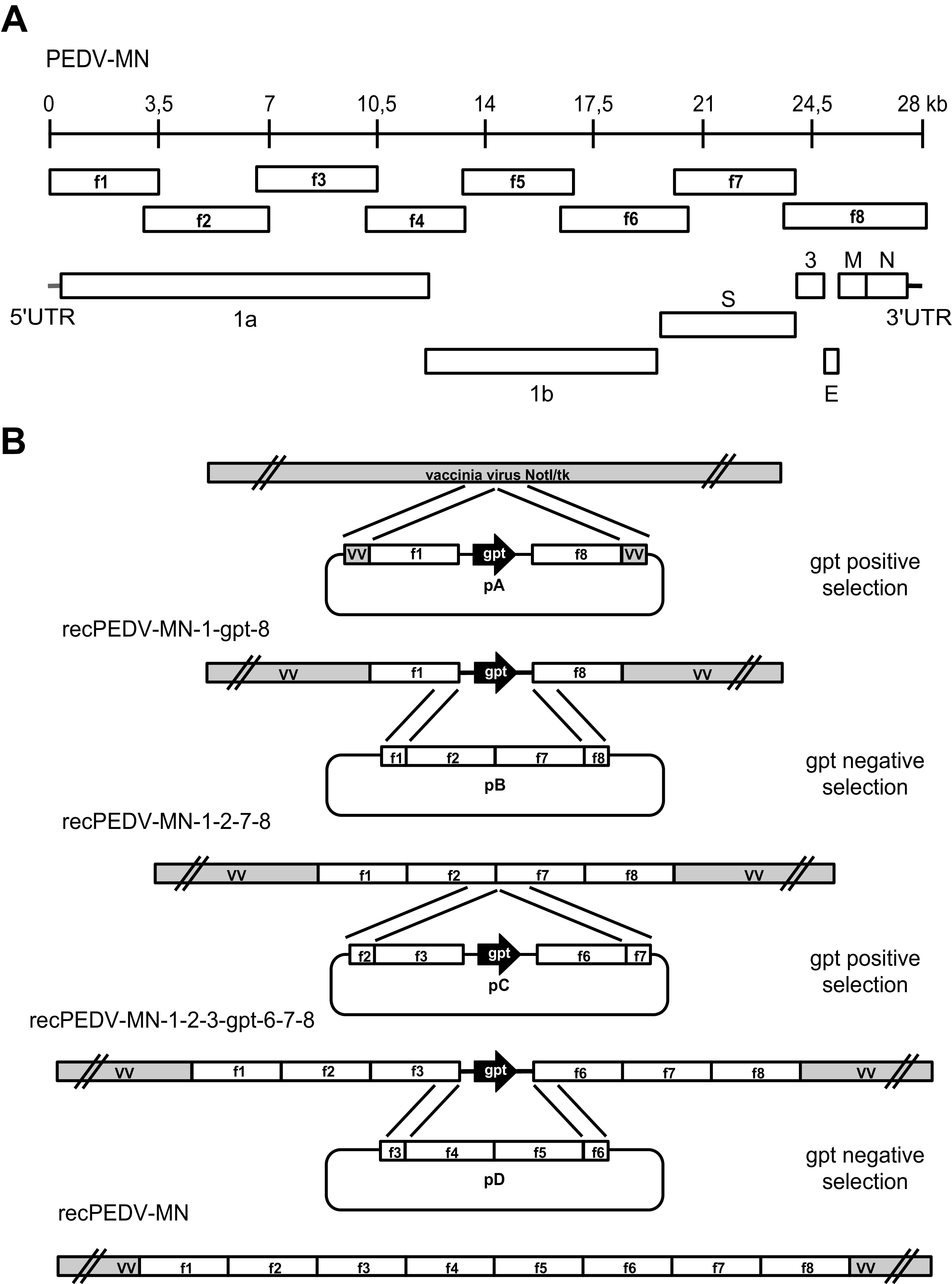
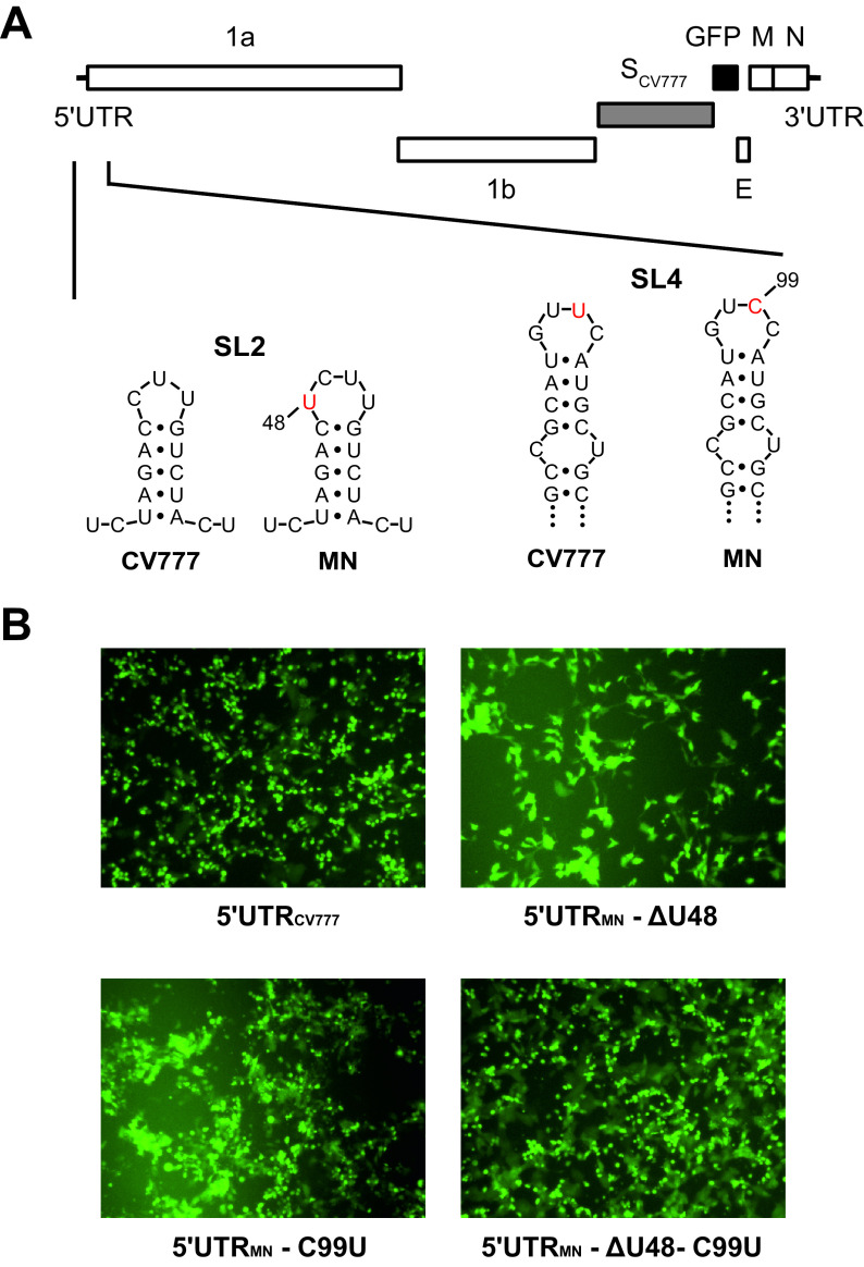
Porcine epidemic diarrhea virus is a swine pathogen that has been responsible for significant animal and economic losses worldwide in recent years. In this manuscript, we report the generation of a reverse genetics system C(RGS) for the highly virulent US PEDV strain Minnesota (PEDV-MN; GenBank accession number KF468752), which was based on the assembly and cloning of synthetic DNA, using vaccinia virus as a cloning vector. Viral rescue was only possible following the substitution of 2 nucleotides within the 5'UTR and 2 additional nucleotides within the spike gene, based on the sequence of the cell culture-adapted strains. Besides displaying a highly pathogenic phenotype in newborn piglets, in comparison with the parental virus, the rescued recombinant PEDV-MN was used to confirm that the PEDV spike gene has an important role in PEDV virulence and that the impact of an intact PEDV ORF3 on viral pathogenicity is modest. Moreover, a chimeric virus with a TGEV spike gene in the PEDV backbone generated with RGS was able to replicate efficiently in vivo and could be readily transmitted between piglets. Although this chimeric virus did not cause severe disease upon the initial infection of piglets, there was evidence of increasing pathogenicity upon transmission to contact piglets. The RGS described in this study constitutes a powerful tool with which to study PEDV pathogenesis and can be used to generate vaccines against porcine enteric coronaviruses. IMPORTANCE PEDV is a swine pathogen that is responsible for significant animal and economic losses worldwide. Highly pathogenic variants can lead to a mortality rate of up to 100% in newborn piglets. The generation of a reverse genetics system for a highly virulent PEDV strain originating from the United States is an important step in phenotypically characterizing PEDV. The synthetic PEDV mirrored the authentic isolate and displayed a highly pathogenic phenotype in newborn piglets. With this system, it was possible to characterize potential viral virulence factors. Our data revealed that an accessory gene (ORF3) has a limited impact on pathogenicity. However, as it is also now known for many coronaviruses, the PEDV spike gene is one of the main determinants of pathogenicity. Finally, we show that the spike gene of another porcine coronavirus, namely, TGEV, can be accommodated in the PEDV genome background, suggesting that similar viruses can emerge in the field via recombination.

Keywords: ORF3; porcine epidemic diarrhea virus; spike gene; synthesized DNA; vaccinia virus reverse genetics.


Conflict of interest statement

The authors declare no conflict of interest.

Figures

FIG 1
FIG 1
Schematic diagram of PEDV genome organization and strategy for the generation of RGS of a highly virulent US PEDV strain (PEDV-MN). (A) The +ssRNA genome of PEDV is represented schematically with a 28 kb scale, the 8 overlapping genome fragments 1 to 8 (f1 to f8) used for cloning the viral cDNA, the 5′ and 3′UTRs, and the ORFs, namely, 1a and 1b, as well as the accessory gene 3 (ORF3), the spike (S), envelope (E), membrane protein (M), and nucleocapsid (N) protein genes. (B) Eight plasmids (p1 to p8) expressing the eight overlapping synthetic cDNA fragments f1 to f8, encompassing the whole PEDV-MN genome, were generated. Subsequently, these plasmids were used to generate four plasmids within the pGPT-1 plasmid backbone, namely, pA, which encoded fragments 1 and 8 upstream and downstream of the gpt gene, respectively; pB, which encoded fragments 2 and 7; pC, which contained fragments 3 and 6 upstream and downstream of the gpt gene, respectively; and pD, which consisted of fragment 5 subcloned into the p4 backbone. These plasmids were then used to introduce the full-length PEDV-MN cDNA into the vaccinia virus NotI/tk backbone in four rounds of vaccinia virus-mediated double recombination, using GPT alternatively as a positive/negative selection marker. These rounds led to the generation of three intermediate constructs, namely, recPEDV-MN-1-gpt-8, recPEDV-MN-1-2-7-8, recPEDV-MN-1-2-3-gpt-6-7-8, and, ultimately, recPEDV-MN.
FIG 2
FIG 2
The rescue of recombinant PEDV-MN-SCV777-ΔORF3-GFP required the repair of the 5′-UTR region. (A) The sequence analysis of 5′UTRMN and 5′UTRCV777 via RACE-PCR disclosed four nucleotide (nt) differences. According to an RNA secondary structure prediction, two of these nucleotides (nt 48 and 99) were located at the tip of stem-loop 2 and stem-loop 4 (SL2 and SL4), respectively. (B) GFP expression was associated with the successful rescue of recombinant viruses containing alterations of the 5′UTRMN, namely, recPEDV-MN-5′UTRCV777-SCV777-ΔORF3-GFP (upper left, 5′UTRCV777), recPEDV-MN-5′UTRMN-ΔU48-SCV777-ΔORF3-GFP (upper right, 5′UTRMN-ΔU48), recPEDV-MN-5′UTRMN-C99U-SCV777-ΔORF3-GFP (lower left, 5′UTRMN-C99U), and recPEDV-5′UTRMN-ΔU48, C99U-SCV777-ΔORF3-GFP (lower right, 5′UTRMN-ΔU48-C99U).
FIG 3
FIG 3
The presence or absence of the ORF3 gene does not significantly impair the recovery of recombinant PEDVs. The sequence analysis and correction of the PEDV-MN spike gene enabled PEDV-MN rescue. (A) Assembly of recPEDV-MN-5′UTRCV777-SCV777-GFP. The last 49 nucleotides from the S gene that include the TRS from ORF3 were inserted downstream of the GFP reporter gene to enable the expression of the ORF3 gene. The GFP expression was associated with the successful rescue of this recombinant virus, as shown on the right. (B) The alignment of selected portions of the amino acid sequence of PEDV-SMN with the corresponding sequences of two highly virulent field strains (PEDV-NPL 2013, PEDV-PC22A) revealed three differences in the receptor binding domain (S1) which are highlighted in red. (C) The replacement of the amino acid at positions 226, 376, and 486 in SMN with the corresponding residues of SPC22A resulted in a green fluorescence signal, indicating the rescue of recPEDV-MN-5′UTRCV777-SMN-F226S, L375F, H486P-GFP. The phenylalanine at position 375 and the proline at 486 (recPEDV-MN-5′UTRCV777-SMN-L375F, H486P-GFP) are essential for virus rescue.
FIG 4
FIG 4
Synthetic recPEDV-MN is highly pathogenic in piglets, and the spike gene plays an important role in PEDV pathogenesis. (A) Clinical scores, body weights, and body temperatures of newborn piglets that were infected with the indicated PEDV isolates and recombinant viruses (recPEDV-MN [n = 6], recPEDV-MN-SCV777 [n = 5], PEDV-NPL 2013 [n = 6], PEDV-CV777 [n = 5]), compared with those of mock-infected piglets (n = 4) until 7 day p.i. Each dot represents a mean value, with error bars showing the standard deviation. (B) Histological and immunohistochemical findings in the jejunum of a representative piglet each at days 4 and 7 p.i., with the indicated viruses shown in comparison to a mock-infected piglet. H&E staining is shown on the left, and anti-PEDVM IHC is shown on the right. Longer bar, 200 μm; shorter bar, 100 μm. (C) Comparison of the jejunal villous length displayed by each of the six piglets from the four groups on days 4 (left) and 7 (right) p.i., in comparison with mock-infected piglets on day 4 p.i. The data were analyzed via a multilevel linear regression model (P < 0.05). One asterisk (*) stands for P < 0.05, three asterisks (***) stand for P < 0.001, and ns stands for a nonsignificant statistical difference.
FIG 5
FIG 5
RNA shedding and RNA distribution in the tissues in the piglets that were infected with synthetic recPEDV-MN resemble the values displayed by PEDV-NPL 2013. (A) Viral RNA shedding in serum, rectal, and oronasal swabs of piglets that were either infected with the indicated viruses or mock-infected. (B) Viral RNA loads in log10 genome equivalents (GE) per mg of tissue samples from primary infected or contact piglets from the four groups that were euthanized at the indicated days p.i. Each dot (panel A) or bar (panel B) represents a mean value, with error bars showing the standard deviation.
FIG 6
FIG 6
The ORF3 gene plays a minor role in PEDV pathogenesis. (A) Clinical scores, body weights and body temperatures of the piglets (n = 6 per group) that were infected with 104 PFU recPEDV-MN-ΔpartORF3 and parental recPEDV-MN, in comparison with mock-infected piglets. (B) Daily viral RNA in log10 GE per 2 mL in serum, rectal, and oronasal swabs of piglets that were infected with recPEDV-MN-ΔpartORF3 and recPEDV-MN, in comparison with mock-infected piglets. (A) Viral RNA load in log10 GE per mg tissue detected in organ samples from piglets on day 4 p.i. with recPEDV-MN-ΔpartORF3 and recPEDV-MN. Each dot (panels A and B) or bar (panel C) represents a mean value, with error bars showing the standard deviation. (D) Histological and immunohistochemical findings in the jejunum of a representative piglet per group. H&E staining is shown on the left, and anti-PEDVM IHC is shown on the right. Longer bar, 200 μm; shorter bar, 100 μm. (E) The comparison of villi length displayed at day 4 p.i. by piglets that were infected with both viruses, in comparison with mock-infected piglets.
FIG 7
FIG 7
A lower dose of infection does not significantly impact the outcome of recPEDV-MN-ΔpartORF3 infection in newborn piglets. (A) Clinical scores, body weights, and body temperatures of the piglets infected with 103 PFU recPEDV-MN-ΔpartORF3 and recPEDV-MN, in comparison with mock-infected piglets. (B) Daily viral RNA in GE per 2 mL in serum, rectal, and oronasal swabs of piglets that were infected with recPEDV-MN-ΔpartORF3 and recPEDV-MN, in comparison with mock-infected piglets. (C) Viral RNA load in log10 GE per mg tissue detected in organ samples from piglets on days 2, 4, and 7 p.i. with recPEDV-MN-ΔpartORF3 and recPEDV-MN. Each dot (panels A and B) or bar (panel C) represents a mean value, with error bars showing the standard deviation. (D) Histological and immunohistochemical findings in the jejunum of a representative piglet per group. H&E staining is shown on the left, and anti-PEDVM IHC is shown on the right. Longer bar, 200 μm; shorter bar, 100 μm. (E) Comparison of villi length displayed at day 4 p.i. by piglets infected with both viruses, in comparison with mock-infected piglets.
FIG 8
FIG 8
A chimeric PEDV/TGEV virus is viable in vitro and in vivo, and it has the potential for increased pathogenicity upon in vivo passaging. (A) A group of nine piglets was infected with recPEDV-MN-STGEV and cohoused with contact piglets (n = 3) at 2 days after infection. The viral RNA loads in log10 GE per mg tissue were determined in several tissue samples from infected piglets at days 2 (n = 3), 4 (n = 3), and 7 p.i. (n = 3). (B) Daily viral RNA loads in log10 GE per 2 mL serum, rectal, and oronasal swabs from infected piglets. (C) Clinical scores, body weights, and body temperatures of infected piglets. Each bar (panel A) or dot (panels B and C) represents a mean value, with error bars showing the standard deviation. (D) Viral RNA loads in log10 GE per mg tissue in the indicated contact piglets (n = 3) at day 5 p.i. (E) Histological and immunohistochemical findings in the jejunum of contact piglets 1 and 2 on day 4 post introduction, in comparison with a representative mock-infected piglet. H&E staining is shown in the top panels, and anti-PEDVM IHC is shown in the bottom panels. Longer bar, 200 μm; shorter bar, 100 μm. (F) Comparison of villi length on day 4 post introduction of contact piglets 1 and 2, in comparison with mock-infected piglets. The data were analyzed via a Kruskal-Wallis H test (P < 0.05). Three asterisks (***) stand for P < 0.001, and ns stands for a nonsignificant statistical difference.

Similar articles

Cited by

References

    1. Jung K, Annamalai T, Lu Z, Saif LJ. 2015. Comparative pathogenesis of US porcine epidemic diarrhea virus (PEDV) strain PC21A in conventional 9-day-old nursing piglets vs. 26-day-old weaned pigs. Vet Microbiol 178:31–40. doi:10.1016/j.vetmic.2015.04.022. - DOI - PMC - PubMed
    1. Stevenson GW, Hoang H, Schwartz KJ, Burrough ER, Sun D, Madson D, Cooper VL, Pillatzki A, Gauger P, Schmitt BJ, Koster LG, Killian ML, Yoon KJ. 2013. Emergence of porcine epidemic diarrhea virus in the United States: clinical signs, lesions, and viral genomic sequences. J Vet Diagn Invest 25:649–654. doi:10.1177/1040638713501675. - DOI - PubMed
    1. De Groot R, Baker SC, Baric R, Enjuanes L, Gorbalenya AE, Holmes KV, Perlman S, Poon LL, Rottier P, Talbot PJ, Woo PC, Ziebuhr J. 2012. Family Coronaviridae. p 806–828. In King AMQ, Carstens EB, Lefkowitz EJ (ed), Virus taxonomy: Ninth Report of the International Committee on Taxonomy of Viruses. Elsevier Academic Press, Amsterdam, NL.
    1. Ziebuhr J, Snijder EJ, Gorbalenya AE. 2000. Virus-encoded proteinases and proteolytic processing in the Nidovirales. J Gen Virol 81:853–879. doi:10.1099/0022-1317-81-4-853. - DOI - PubMed
    1. Thiel V, Herold J, Schelle B, Siddell SG. 2001. Infectious RNA transcribed in vitro from a cDNA copy of the human coronavirus genome cloned in vaccinia virus. J Gen Virol 82:1273–1281. doi:10.1099/0022-1317-82-6-1273. - DOI - PubMed

Publication types

Substances

LinkOut - more resources