. 2023 Jun 1;7(6):508-527.
doi: 10.4049/immunohorizons.2300034.

Highly Networked SARS-CoV-2 Peptides Elicit T Cell Responses with Enhanced Specificity

Affiliations

Highly Networked SARS-CoV-2 Peptides Elicit T Cell Responses with Enhanced Specificity

Gabriel Duette et al. Immunohorizons. .

Abstract

Identifying SARS-CoV-2-specific T cell epitope-derived peptides is critical for the development of effective vaccines and measuring the duration of specific SARS-CoV-2 cellular immunity. In this regard, we previously identified T cell epitope-derived peptides within topologically and structurally essential regions of SARS-CoV-2 spike and nucleocapsid proteins by applying an immunoinformatics pipeline. In this study, we selected 30 spike- and nucleocapsid-derived peptides and assessed whether these peptides induce T cell responses and avoid major mutations found in SARS-CoV-2 variants of concern. Our peptide pool was highly specific, with only a single peptide driving cross-reactivity in people unexposed to SARS-COV-2, and immunogenic, inducing a polyfunctional response in CD4+ and CD8+ T cells from COVID-19 recovered individuals. All peptides were immunogenic and individuals recognized broad and diverse peptide repertoires. Moreover, our peptides avoided most mutations/deletions associated with all four SARS-CoV-2 variants of concern while retaining their physicochemical properties even when genetic changes are introduced. This study contributes to an evolving definition of individual CD4+ and CD8+ T cell epitopes that can be used for specific diagnostic tools for SARS-CoV-2 T cell responses and is relevant to the development of variant-resistant and durable T cell-stimulating vaccines.


Conflict of interest statement

The authors have no financial conflicts of interest.

Figures

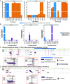
FIGURE 1.
FIGURE 1.
T cell response induced by highly networked SARS-CoV-2 peptide pool. Selected highly networked (HN) SARS-CoV-2 peptides were combined in a single pool and tested in PBMCs from recovered COVID-19 participants. Alternatively, cells were stimulated with commercial spike (S) or nucleocapsid (NC) overlapping peptide pools or PHA. (AC) Cytokine production was assessed by FluoroSpot (A and B) and flow cytometry (C). (A) Representative FluoroSpot showing IFN-γ (green spots) and IL-2 (red spots) production by PBMCs from three recovered COVID-19 donors. (B) Quantification of spot-forming units (SFU) per 106 cells for IFN-γ, IL-2, or dual-secreting cells. Each data point represents a single donor (n = 29) with bars showing the median. Green dots represent values obtained from recovered COVID-19 participants infected with the Delta variant of SARS-CoV-2. All others were infected with the ancestral strain. Statistical significance was assessed using Friedman’s test, and p values were not significant (IFN-γ, p = 0.147; IL-2, p = 0.200; IFN-γ/IL-2, p = 0.132). (C) Representative intracellular flow cytometry data showing effector cytokine production and degranulation in CD4+ T cells and CD8+ T cells expanded in vitro with the HN SARS-CoV-2 peptide pool. Pie charts depict the average distribution of mono-, bi-, tri-, and tetrafunctional SARS-CoV-2–specific CD4+ and CD8+ T cells.
FIGURE 2.
FIGURE 2.
T cell response to individual NC and S-derived HN SARS-CoV-2 peptides. Cytokine production by PBMCs from recovered COVID-19 participants exposed to HN SARS-CoV-2 individual peptides was assessed by FluoroSpot. (A and B) Quantification of spot-forming units (SFU) per 106 cells secreting IFN-γ and IL-2 after stimulation with MHC class II (A)– and MHC class I (B)–restricted peptides or commercial S and NC overlapping peptide pools. Each data point represents a single donor (n = 32 for MHC class II and n = 7 for MHC class I) and bars show the median. Green dots represent values obtained from a recovered COVID-19 participant infected with the Delta variant of SARS-CoV-2.
FIGURE 3.
FIGURE 3.
Polyfunctionality of the T cell response to individual NC and S-derived HN SARS-CoV-2 peptides. T cell polyfunctionality analysis after in vitro expansion with SARS CoV-2 peptides. (A and B) Representative dot plots showing intracellular cytokine production and degranulation level in CD4+ T cells (A) and CD8+ T cells (B) induced by individual HN SARS-CoV-2 peptides. (C and D) Pies depict the average distribution across n reactive donors of mono-, bi-, tri-, and tetrafunctional cells within SARS-CoV-2–specific CD4+ T cells (C) and CD8+ T cells (D).
FIGURE 4.
FIGURE 4.
Cross-reactive T cell response to HN SARS-CoV-2 peptides. Cytokine production by PBMCs from donors unexposed to SARS-CoV-2 (pre–COVID-19) stimulated with SARS-CoV-2 peptides was assessed by FluoroSpot. (A) Quantification of spot-forming units (SFU) per 106 cells secreting IFN-γ and IL-2 after stimulation with SARS-CoV-2 peptide pools. Data obtained from COVID-19 recovered donors are shown for comparison. Green dots represent values obtained from recovered COVID-19 participants infected with the Delta variant of SARS-CoV-2. All others were infected with the ancestral strain. (B) Quantification of SFU per 106 cells of IFN-γ and IL-2 and after stimulation with individual MHC class I (A)– and MHC class II (B)–restricted peptides. Each dot color represents a single donor. (C) Quantification of SFU per 106 cells of IFN-γ and IL-2 after stimulation with SARS-CoV-2 peptide pools. Each data point represents a single donor and bars show the median. Statistical significance was assessed using the Mann–Whitney test. * p ≤ 0.05, ** p ≤ 0.01, ***p ≤ 0.001, ****p ≤ 0.0001; ns, not significant.
FIGURE 5.
FIGURE 5.
Comparison of mutations within SARS-CoV-2 S protein associated with VOCs and T cell escape to the peptides selected for ex vivo assessment. (A and B) The proportion of S protein sequences containing mutations that define Alpha, Beta, and Delta VOCs (A) and the proportion of Alpha, Beta, and Delta VOC S protein sequences containing known T cell escape mutations (B) are shown. The proportion of S protein sequences with ambiguous amino acid residues (X) at the mutation sites is marked by blue bars (A and B). The threshold for determining the most significant mutations for Alpha, Beta, and Delta VOCs is denoted by the dotted line (≥1%; A and B). (C and D) All of the mutations were compared with the peptides selected from S subunit 1 (C) and subunit 2 (D). The 25 peptides are indicated by dark (≥12-mer) and light (9-mer) maroon bars. Detailed legends are provided within the figure panels (C and D).
FIGURE 6.
FIGURE 6.
Comparison of mutations within SARS-CoV-2 S protein associated with Omicron VOC and its sublineages to the peptides selected for ex vivo assessment. (A and B) The mutations that define the Omicron SARS-CoV-2 variants were compared with the selected peptides for S subunit 1 (A) and subunit 2 (B) protein regions.
FIGURE 7.
FIGURE 7.
Physicochemical properties of the six S-derived peptides with and without major mutations and deletions found within Alpha, Beta, and Omicron VOCs. (AD) The mean hydrophobicity (A), isoelectric point (B), molecular mass (C), and geometric distance (D) of each S-derived peptide with or without major mutations and deletions associated with Alpha, Beta, and Omicron VOCs are shown. The units for molecular mass and geometric distance are Daltons and angstroms, respectively. The six S-derived peptides include those that contained mutations/deletions that were found within ≥1% of Alpha and Beta variant sequences, or those that contained at least 1 of 32 mutations associated with the Omicron variant. The peptide sequences that do not contain the mutations/deletions are underlined. The mutations/deletions are indicated by bolded red texts in the peptide sequences.
FIGURE 8.
FIGURE 8.
Comparison of mutations within SARS-CoV-2 NC protein associated with VOCs to the peptides selected for ex vivo assessment. (A) The mutations that appear within at least 1% of the NC protein sequences derived from Alpha, Beta, and Delta VOCs were indicated and compared with the seven peptides selected for ex vivo assay. (B) The proportion of NC protein sequences derived from Alpha and Delta VOCs that contained L139, the L139F mutation, ambiguous amino acid residues, and other mutations is shown. (C) The mutations associated with NC protein sequences of the Omega and its sublineages were compared with the peptides selected for ex vivo assay.

Similar articles

Cited by

References

    1. Sette, A., Crotty S.. 2021. Adaptive immunity to SARS-CoV-2 and COVID-19. Cell 184: 861–880. - PMC - PubMed
    1. Bastard, P., Rosen L. B., Zhang Q., Michailidis E., Hoffmann H. H., Zhang Y., Dorgham K., Philippot Q., Rosain J., Béziat V., et al. COVID Human Genetic Effort . 2020. Autoantibodies against type I IFNs in patients with life-threatening COVID-19. Science 370: eabd4585. - PMC - PubMed
    1. Galani, I. E., Rovina N., Lampropoulou V., Triantafyllia V., Manioudaki M., Pavlos E., Koukaki E., Fragkou P. C., Panou V., Rapti V., et al. . 2021. Untuned antiviral immunity in COVID-19 revealed by temporal type I/III interferon patterns and flu comparison. Nat. Immunol. 22: 32–40. - PubMed
    1. Hadjadj, J., Yatim N., Barnabei L., Corneau A., Boussier J., Smith N., Péré H., Charbit B., Bondet V., Chenevier-Gobeaux C., et al. . 2020. Impaired type I interferon activity and inflammatory responses in severe COVID-19 patients. Science 369: 718–724. - PMC - PubMed
    1. Tan, A. T., Linster M., Tan C. W., Le Bert N., Chia W. N., Kunasegaran K., Zhuang Y., Tham C. Y. L., Chia A., Smith G. J. D., et al. . 2021. Early induction of functional SARS-CoV-2-specific T cells associates with rapid viral clearance and mild disease in COVID-19 patients. Cell Rep. 34: 108728. - PMC - PubMed

Publication types

Supplementary concepts