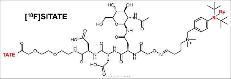
Next-generation PET/CT imaging in meningioma-first clinical experiences using the novel SSTR-targeting peptide [18F]SiTATE
- PMID: 37358620
- PMCID: PMC10541820
- DOI: 10.1007/s00259-023-06315-z
Next-generation PET/CT imaging in meningioma-first clinical experiences using the novel SSTR-targeting peptide [18F]SiTATE
Erratum in
-
Correction to: Next‑generation PET/CT imaging in meningioma-first clinical experiences using the novel SSTR‑targeting peptide [18F]SiTATE.Eur J Nucl Med Mol Imaging. 2023 Nov;50(13):4115. doi: 10.1007/s00259-023-06409-8. Eur J Nucl Med Mol Imaging. 2023. PMID: 37642707 Free PMC article. No abstract available.
Abstract
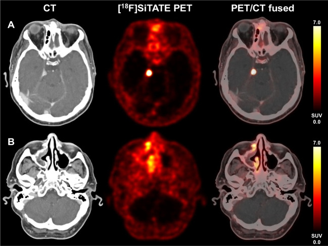
Background: Somatostatin-receptor (SSTR)-targeted PET/CT provides important clinical information in addition to standard imaging in meningioma patients. [18F]SiTATE is a novel, 18F-labeled SSTR-targeting peptide with superior imaging properties according to preliminary data. We provide the first [18F]SiTATE PET/CT data of a large cohort of meningioma patients.
Methods: Patients with known or suspected meningioma undergoing [18F]SiTATE PET/CT were included. Uptake intensity (SUV) of meningiomas, non-meningioma lesions, and healthy organs were assessed using a 50% isocontour volume of interest (VOI) or a spherical VOI, respectively. Also, trans-osseous extension on PET/CT was assessed.
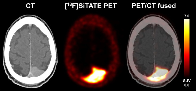
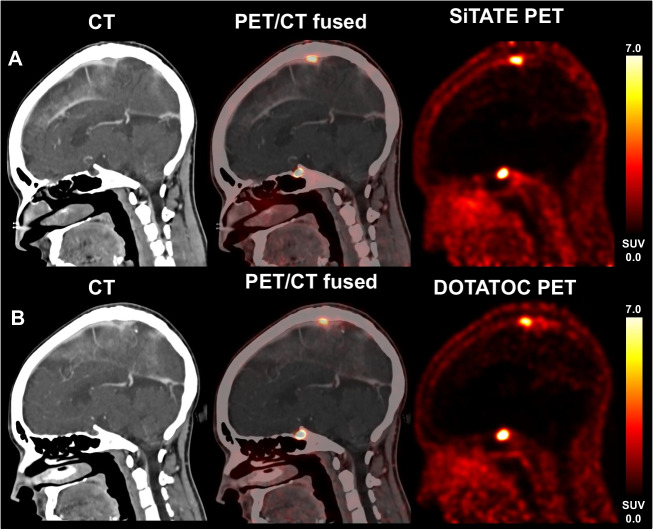
Results: A total of 107 patients with 117 [18F]SiTATE PET/CT scans were included. Overall, 231 meningioma lesions and 61 non-meningioma lesions (e.g., post-therapeutic changes) were analyzed. Physiological uptake was lowest in healthy brain tissue, followed by bone marrow, parotid, and pituitary (SUVmean 0.06 ± 0.04 vs. 1.4 ± 0.9 vs. 1.6 ± 1.0 vs. 9.8 ± 4.6; p < 0.001). Meningiomas showed significantly higher uptake than non-meningioma lesions (SUVmax 11.6 ± 10.6 vs. 4.0 ± 3.3, p < 0.001). Meningiomas showed significantly higher uptake than non-meningioma lesions (SUVmax 11.6±10.6 vs. 4.0±3.3, p<0.001). 93/231 (40.3%) meningiomas showed partial trans-osseous extension and 34/231 (14.7%) predominant intra-osseous extension. 59/231 (25.6%) meningioma lesions found on PET/CT had not been reported on previous standard imaging.
Conclusion: This is the first PET/CT study using an 18F-labeled SSTR-ligand in meningioma patients: [18F]SiTATE provides extraordinary contrast in meningioma compared to healthy tissue and non-meningioma lesions, which leads to a high detection rate of so far unknown meningioma sites and osseous involvement. Having in mind the advantageous logistic features of 18F-labeled compared to 68Ga-labeled compounds (e.g., longer half-life and large-badge production), [18F]SiTATE has the potential to foster a widespread use of SSTR-targeted imaging in neuro-oncology.
Keywords: Meningioma; PET; SiTATE; Somatostatin receptor (SSTR).
© 2023. The Author(s).
Conflict of interest statement
JCT received research grants from Novocure and Munich Surgical Imaging and lecture honoraria from Seagen. NLA received research grants from Novocure and honoraria for consultation or advisory board participation from Novartis and Telix. All other authors do not declare any potential conflict of interest.
Figures