

Diffuse pancreatic parenchymal atrophy, an imaging finding predictive of the development of pancreatic ductal adenocarcinoma: A case-control study
- PMID: 37359111
- PMCID: PMC10290266
- DOI: 10.1002/jgh3.12930
Diffuse pancreatic parenchymal atrophy, an imaging finding predictive of the development of pancreatic ductal adenocarcinoma: A case-control study
Abstract
Background and aim: Pancreatic ductal adenocarcinoma (PDAC) is a lethal cancer, partly because its early detection is difficult. This study aimed to identify computed tomography (CT) findings associated with PDAC prior to diagnosis.
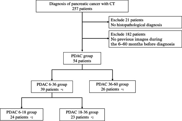
Methods: Past CT images were retrospectively collected from the PDAC group (n = 54) and the control group (n = 90). The following imaging findings were compared: pancreatic mass, main pancreatic duct (MPD) dilatation with or without cutoff, cyst, chronic pancreatitis with calcification, partial parenchymal atrophy (PPA), and diffuse parenchymal atrophy (DPA). In the PDAC group, CT findings were examined during the pre-diagnostic period and 6-36 months and 36-60 months before diagnosis. Multivariate analyses were performed using logistic regression.
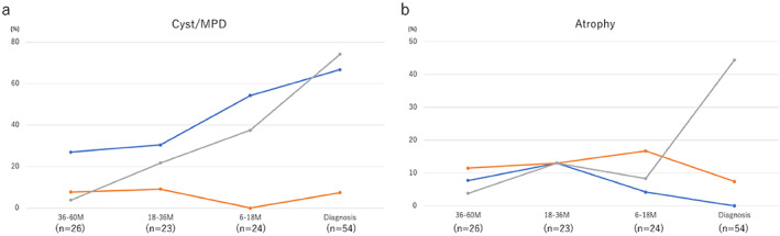
Results: MPD dilatation with cutoff (P < 0.0001) and PPA (P = 0.023) were identified as significant imaging findings 6-36 months before diagnosis. DPA was identified as a novel imaging finding at 6-36 months (P = 0.003) and 36-60 months (P = 0.009) before diagnosis.
Conclusion: DPA, MPD dilatation with cutoff, and PPA were identified as imaging findings associated with pre-diagnostic PDAC.
Keywords: computed tomography; main pancreatic duct; pancreatic cancer; pancreatic ductal adenocarcinoma; pancreatic parenchymal atrophy.
© 2023 The Authors. JGH Open published by Journal of Gastroenterology and Hepatology Foundation and John Wiley & Sons Australia, Ltd.
Figures
 , Cyst;
  , MPD dilatation without cutoff;
  , MPD dilatation with cutoff. (b):
  , DPA;
  , FPA;
  , UPA.

References
-
- Bray F, Ferlay J, Soerjomataram I, Siegel RL, Torre LA, Jemal A. Global cancer statistics 2018: GLOBOCAN estimates of incidence and mortality worldwide for 36 cancers in 185 countries. CA Cancer J. Clin. 2018; 68: 394–424. - PubMed
-
- Egawa S, Toma H, Ohigashi H et al. Japan Pancreatic Cancer Registry; 30th year anniversary: Japan Pancreas Society. Pancreas. 2012; 41: 985–92. - PubMed
-
- Yamaguchi K, Okusaka T, Shimizu K et al. Clinical practice guidelines for pancreatic cancer 2016 from the Japan Pancreas Society: a synopsis. Pancreas. 2017; 46: 595–604. - PubMed
-
- Kanno A, Masamune A, Hanada K et al. Multicenter study of early pancreatic cancer in Japan. Pancreatology. 2018; 18: 61–7. - PubMed
-
- Vasen HFA, Boekestijn B, Ibrahim IS et al. Dilatation of the main pancreatic duct as first manifestation of small pancreatic ductal adenocarcinomas detected in a hereditary pancreatic cancer surveillance program. HPB (Oxford). 2019; 21: 1371–5. - PubMed
LinkOut - more resources
Full Text Sources
Miscellaneous
