

Small bowel capsule endoscopy examination and open access database with artificial intelligence: The SEE-artificial intelligence project
- PMID: 37359150
- PMCID: PMC10288072
- DOI: 10.1002/deo2.258
Small bowel capsule endoscopy examination and open access database with artificial intelligence: The SEE-artificial intelligence project
Abstract
Objectives: Artificial intelligence (AI) may be practical for image classification of small bowel capsule endoscopy (CE). However, creating a functional AI model is challenging. We attempted to create a dataset and an object detection CE AI model to explore modeling problems to assist in reading small bowel CE.
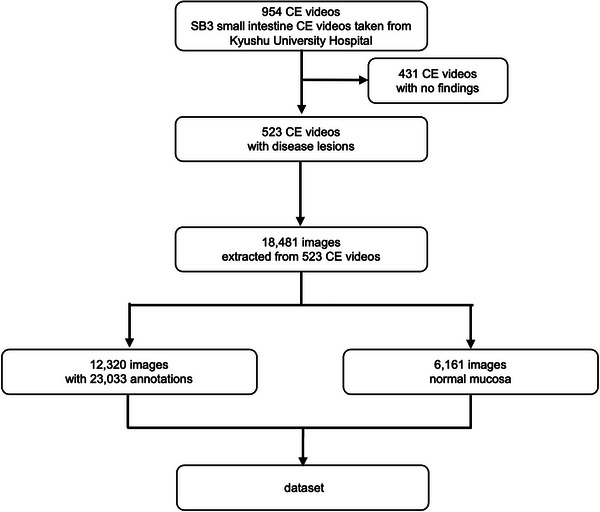
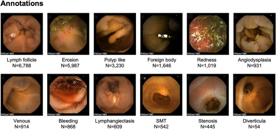
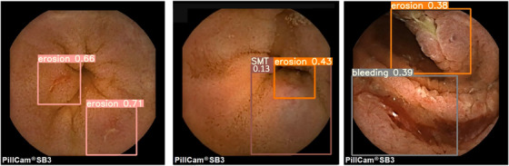
Methods: We extracted 18,481 images from 523 small bowel CE procedures performed at Kyushu University Hospital from September 2014 to June 2021. We annotated 12,320 images with 23,033 disease lesions, combined them with 6161 normal images as the dataset, and examined the characteristics. Based on the dataset, we created an object detection AI model using YOLO v5 and we tested validation.
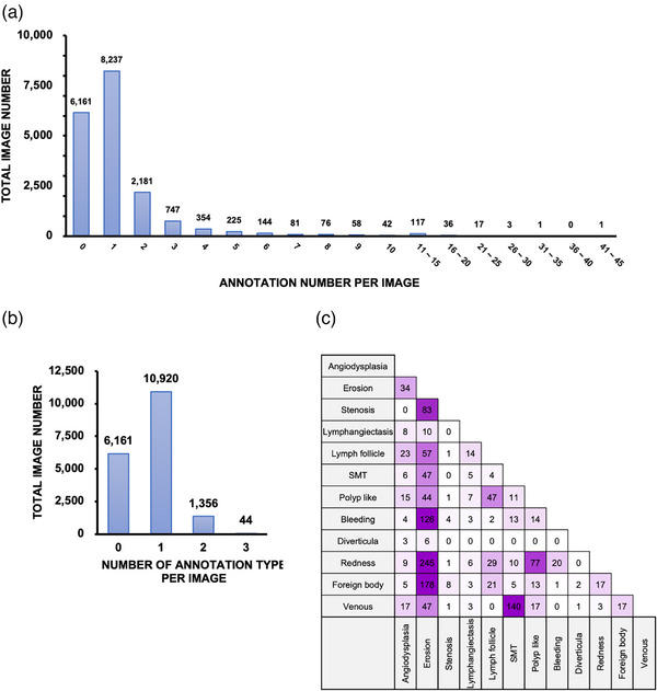
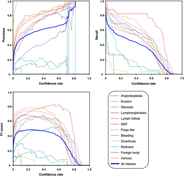
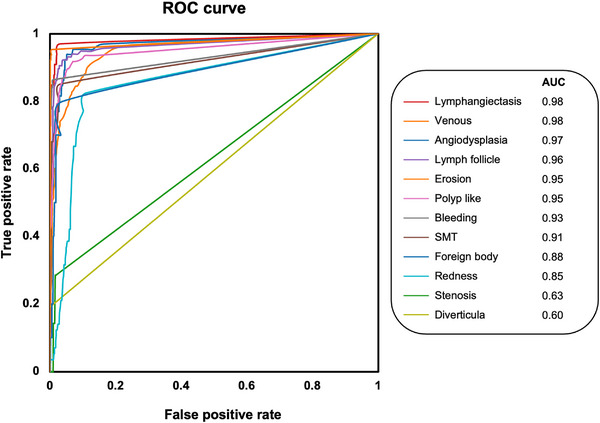
Results: We annotated the dataset with 12 types of annotations, and multiple annotation types were observed in the same image. We test validated our AI model with 1396 images, and sensitivity for all 12 types of annotations was about 91%, with 1375 true positives, 659 false positives, and 120 false negatives detected. The highest sensitivity for individual annotations was 97%, and the highest area under the receiver operating characteristic curve was 0.98, but the quality of detection varied depending on the specific annotation.
Conclusions: Object detection AI model in small bowel CE using YOLO v5 may provide effective and easy-to-understand reading assistance. In this SEE-AI project, we open our dataset, the weights of the AI model, and a demonstration to experience our AI. We look forward to further improving the AI model in the future.
Keywords: artificial intelligence; capsule endoscopy; diagnostic imaging; gastrointestinal tract; intestine small.
© 2023 The Authors. DEN Open published by John Wiley & Sons Australia, Ltd on behalf of Japan Gastroenterological Endoscopy Society.
Conflict of interest statement
None.
Figures
References
-
- Iddan G, Meron G, Glukhovsky A, Swain P. Wireless capsule endoscopy. Nature 2000; 405: 417. - PubMed
-
- Hosoe N, Takabayashi K, Ogata H, Kanai T. Capsule endoscopy for small‐intestinal disorders: Current status. Dig Endosc 2019; 31: 498–507. - PubMed
-
- Enns RA, Hookey L, Armstrong D et al. Clinical practice guidelines for the use of video capsule endoscopy. Gastroenterology 2017; 152: 497–514. - PubMed
-
- Cave DR, Hakimian S, Patel K. Current controversies concerning capsule endoscopy. Dig Dis Sci 2019; 64: 3040–7. - PubMed
-
- Aoki T, Yamada A, Aoyama K et al. Clinical usefulness of a deep learning‐based system as the first screening on small‐bowel capsule endoscopy reading. Dig Endosc 2020; 32: 585–91. - PubMed
LinkOut - more resources
Full Text Sources