. 2023 Jun 3:33:1-14.
doi: 10.1016/j.omtn.2023.05.024. eCollection 2023 Sep 12.

Engineering B cells with customized therapeutic responses using a synthetic circuit

Affiliations

Engineering B cells with customized therapeutic responses using a synthetic circuit

Audrey Page et al. Mol Ther Nucleic Acids. .

Abstract

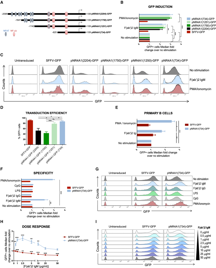
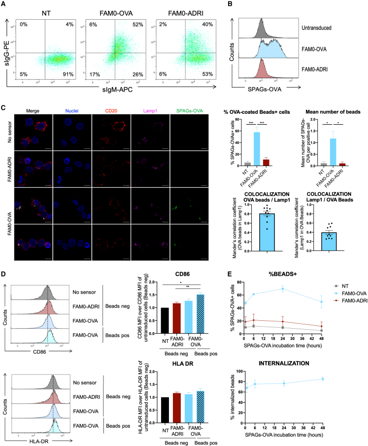
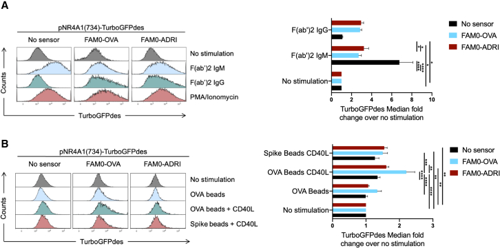
The expansion of genetic engineering has brought a new dimension for synthetic immunology. Immune cells are perfect candidates because of their ability to patrol the body, interact with many cell types, proliferate upon activation, and differentiate in memory cells. This study aimed at implementing a new synthetic circuit in B cells, allowing the expression of therapeutic molecules in a temporally and spatially restricted manner that is induced by the presence of specific antigens. This should enhance endogenous B cell functions in terms of recognition and effector properties. We developed a synthetic circuit encoding a sensor (a membrane-anchored B cell receptor targeting a model antigen), a transducer (a minimal promoter induced by the activated sensor), and effector molecules. We isolated a 734-bp-long fragment of the NR4A1 promoter, specifically activated by the sensor signaling cascade in a fully reversible manner. We demonstrate full antigen-specific circuit activation as its recognition by the sensor induced the activation of the NR4A1 promoter and the expression of the effector. Overall, such novel synthetic circuits offer huge possibilities for the treatment of many pathologies, as they are completely programmable; thus, the signal-specific sensors and effector molecules can be adapted to each disease.

Keywords: B cells; MT: Delivery Strategies; autologous; cell therapy; personalized medicine; reprogramming; synthetic circuits.


Conflict of interest statement

The authors declare no conflict of interest.

Figures

None
Graphical abstract
Figure 1
Figure 1
Isolation and characterization of a small ectopic BCR-inducible promoter construct (A) Schematic representation of the NR4A1 reporter constructs with putative binding domains of the NF-κB and NFAT transcription factors. Different LVs encoding a reporter gene (GFP) with several promoter sizes (2,204, 1,750, 1,251, or 7,34 bp) have been constructed. (B) GFP induction of the reporter constructs after 24 h of BCR stimulation. BJAB cells were transduced with LVs encoding the reporter constructs (NR4A1 or SFFV as a constitutive control) before stimulation with F(ab’)2 IgM or PMA (combined with ionomycin for 24 h). The median GFP expression of GFP+ cells after stimulation was normalized by the non-stimulated condition. (C) Representative flow cytometry histograms of GFP expression are presented for one replicate. (D) Transduction efficiency of BJAB cells by the reporter constructs. The percentage of GFP+ cells after transduction was assessed for each construct 4 days after transduction. (E) Induction of the promoter in primary human B cells. Primary human B cells isolated from peripheral blood were transduced with LVs encoding the GFP under the 734-bp inducible reporter or the SFFV constitutive reporter before 24-h stimulation with F(ab’)2 Ig or PMA combined with ionomycin. GFP median expression was assessed by flow cytometry in GFP+ cells. The induction fold was computed as the median GFP intensity in GFP+ cells after stimulation over the median of non-stimulated GFP+ cells. (F) Specificity of the induction through the BCR. BJAB cells were transduced with LVs encoding the reporter constructs (734-bp NR4A1 or SFFV as a constitutive control) before stimulation with F(ab’)2 IgM, F(ab’)2 IgG, LPS, or CpG or PMA combined with ionomycin for 24 h. The median GFP expression of GFP+ cells after stimulation was normalized by the non-stimulated condition. (G) Representative flow cytometry histograms are presented for one replicate. (H) Dose-response of the 734-bp NR4A1 reporter construct with increasing amounts of F(ab’)2 IgM. BJAB cells were transduced with LVs encoding the 734-bp inducible reporter or the SFFV constitutive reporter before 24 h of stimulation with F(ab’)2 IgM. GFP median expression of DAPI- GFP+ cells relative to unstimulated cells was assessed. I. Representative histograms of GFP expression are presented. Data in (B, D, E, F and H) are representative of four independent experiments (n = 4, ANOVA 2 and multiple t-tests with Sidak Bonferroni correction [alpha = 0.05]). Error bars represent SEM. p values under 0.05 were considered statistically significant and the following denotations were used: ∗∗∗, p<0.001; ∗∗, p<0.01; ∗, p<0.05.
Figure 2
Figure 2
Kinetics characterization of the inducible 734-bp NR4A1 promoter (A) Scheme of the kinetics characterization set-up. (B) Kinetics of induction of the promoter. BJAB cells were transduced with LVs encoding the TurboGFPdes under the 734-bp inducible reporter before 0 to 24 h of stimulation with F(ab’)2 IgM or PMA combined with ionomycin. TurboGFPdes median expression in each condition (after the indicated stimulation duration) was assessed and normalized by TurboGFPdes median of unstimulated cells (n = 3). (C) Kinetics of extinction of the promoter. Transduced BJAB cells were stimulated for 0, 1, 4, 8, and 24 h with F(ab’)2 IgM or PMA combined with ionomycin. Cells were then washed three times and kept in culture. TurboGFPdes median expression was assessed at days 1, 3, and 6 after washing and normalized by TurboGFPdes median of unstimulated cells (n = 3). (D) Reversibility of the inducible promoter. Reversibility of pNR4A1(734)-responsive TurboGFPdes expression was assessed by culturing transduced BJAB cells while alternating 8 h of stimulation (gray) and 88 h of rest (white) three times with F(ab’)2 IgM. TurboGFPdes median expression was assessed before and after stimulation and normalized by TurboGFPdes median of unstimulated cells (n = 3). Error bars represent SEM.
Figure 3
Figure 3
Design and validation of a synthetic sensor (A) Validation of sensor expression. BJAB cells (IgM+, IgG) were transduced with LVs encoding sensors (membrane anchored IgG) recognizing either OVA (FAM0-OVA) or the HbS glycoprotein of HBV (FAM0-ADRI) 5 days before staining of IgG by flow cytometry. (B and C) Specific recognition of OVA by membrane-anchored BCR directed against OVA. BJAB cells were transduced with LVs encoding membrane-anchored BCR targeting either OVA or HbS. Five days after transduction, these cells were incubated for 24 h with OVA-coated fluorescent beads. Binding of fluorescent beads was assessed by flow cytometry (B) or immunofluorescence (C). For immunofluorescence, transduced cells were incubated overnight with OVA-coated fluorescent beads before being washed, fixed, and permeabilized for staining with an anti-Lamp1 antibody and Hoechst. Images were acquired on a confocal microscope. Scale bar, 10 μm. Four fields for each condition were quantified to assess the percentage of cells with beads as well as the median number of beads per cells. The Mander’s correlation coefficients of OVA beads and Lamp1 colocalization were computed. (D) Signaling and cell activation after SPAGs-OVA binding. BJAB cells transduced with membrane BCRs recognizing HbS or OVA were incubated for 24 h with SPAGs-OVA before staining with an anti-CD86 or an anti-HLA-DR antibody. One representative overlay is presented along with the median fluorescence intensity quantified from three experiments for both markers (ANOVA1 and multiple comparison with Tukey correction, n = 3). (E) Internalization of SPAGs-OVA specifically by the OVA sensors. BJAB cells transduced with membrane BCRs recognizing HbS or OVA were incubated for different times (1 h, 6 h, 24 h, or 48 h) with SPAGs-OVA before staining with an anti-OVA antibody (surface staining). The percentage of cells positive for SPAGs-OVA as well as the percentage of internalization computed as the percentage of SPAGs-OVA+/OVA-cells over the percentage of SPAGs-OVA+ cells are presented (n = 3). p values under 0.05 were considered statistically significant and the following denotations were used: ∗∗∗, p<0.001; ∗∗, p<0.01; ∗, p<0.05.
Figure 4
Figure 4
Antigen-specific activation of the synthetic circuit BJAB cells were transduced with LVs encoding pNR4A1(734)-TurboGFPdes 1 week before transduction with LVs encoding a membrane-anchored BCR directed against OVA (FAM0-OVA) or against HBs (FAM0-ADRI). Double transduced cells were then stimulated either with F(ab’)2 directed against IgM or IgG, or PMA/ionomycin as a control (A) or OVA/Spike-RBD-coated beads with or without CD40L (B) for 24 h before detection of TurboGFPdes fluorescence by flow cytometry. One representative overlay is presented along with the median fluorescence intensity quantified from five or six experiments (ANOVA 2 post hoc comparison with Tukey correction). p values under 0.05 were considered statistically significant and the following denotations were used: ∗∗∗, p<0.001; ∗∗, p<0.01; ∗, p<0.05.
Figure 5
Figure 5
All-in-one vectors encoding a self-amplifying circuit (A) Structure of self-amplifying vector. The inducible pNR4A1-734bp promoter fragment drives both the TurboGFPdes and the FAM0-OVA sensor transgenes with a T2A sequence in between. (B and E) Self-amplifying vector induction of effector expression in transduced BJAB cells after 24 h (B) or 48 h (E) stimulation with F(ab’)2 IgM or F(ab’)2 IgG. (C and F) Self-amplifying vector induction of sensor expression in transduced BJAB cells after 24 h (C) or 48 h (F) stimulation with F(ab’)2 IgM or F(ab’)2 IgG. The fold change of the mRNAs encoding the sensor was assessed by RT-qPCR after stimulation. (D and G) Self-amplifying vector induction of effector expression in transduced BJAB cells stimulated with OVA/Spike RBD-coated beads with or without CD40L for 24 h (D) or 48 h (G) before detection of TurboGFPdes fluorescence by flow cytometry. The median expression of TurboGFPdes was assessed by flow cytometry normalized by the median of unstimulated cells. ANOVA 2 post hoc comparison with Tukey correction (n = 5/6 for [B, D, E, G] and n = 3 for [C, F]). Error bars represent SEM. p values under 0.05 were considered statistically significant and the following denotations were used: ∗∗∗, p<0.001; ∗∗, p<0.01; ∗, p<0.05.

References

    1. Sedlmayer F., Aubel D., Fussenegger M. Synthetic gene circuits for the detection, elimination and prevention of disease. Nat. Biomed. Eng. 2018;2:399–415. doi: 10.1038/s41551-018-0215-0. - DOI - PubMed
    1. Roybal K.T., Lim W.A. Synthetic immunology: hacking immune cells to expand their therapeutic capabilities. Annu. Rev. Immunol. 2017;35:229–253. doi: 10.1146/annurev-immunol-051116-052302. - DOI - PMC - PubMed
    1. Bugatti S., Vitolo B., Caporali R., Montecucco C., Manzo A. B cells in rheumatoid arthritis: from pathogenic players to disease biomarkers. BioMed Res. Int. 2014;2014:681678–681714. doi: 10.1155/2014/681678. - DOI - PMC - PubMed
    1. Yuen G.J., Demissie E., Pillai S. B lymphocytes and cancer: a love–hate relationship. Trends Cancer. 2016;2:747–757. doi: 10.1016/j.trecan.2016.10.010. - DOI - PMC - PubMed
    1. Chabannon C., Bouabdallah R., Fürst S., Granata A., Saillard C., Vey N., Mokart D., Fougereau E., Lemarie C., Mfarrej B., et al. CAR-T cells : lymphocytes exprimant un récepteur chimérique à l’antigène. Rev. Med. Interne. 2019;40:545–552. doi: 10.1016/j.revmed.2018.12.002. - DOI - PubMed