Review
. 2023 Jun 8:101718.
doi: 10.1016/j.cocis.2023.101718. Online ahead of print.

Engineering Innovative Interfaces for Point-of-Care Diagnostics

Affiliations
Review

Engineering Innovative Interfaces for Point-of-Care Diagnostics

Damon T Burrow et al. Curr Opin Colloid Interface Sci. .

Abstract

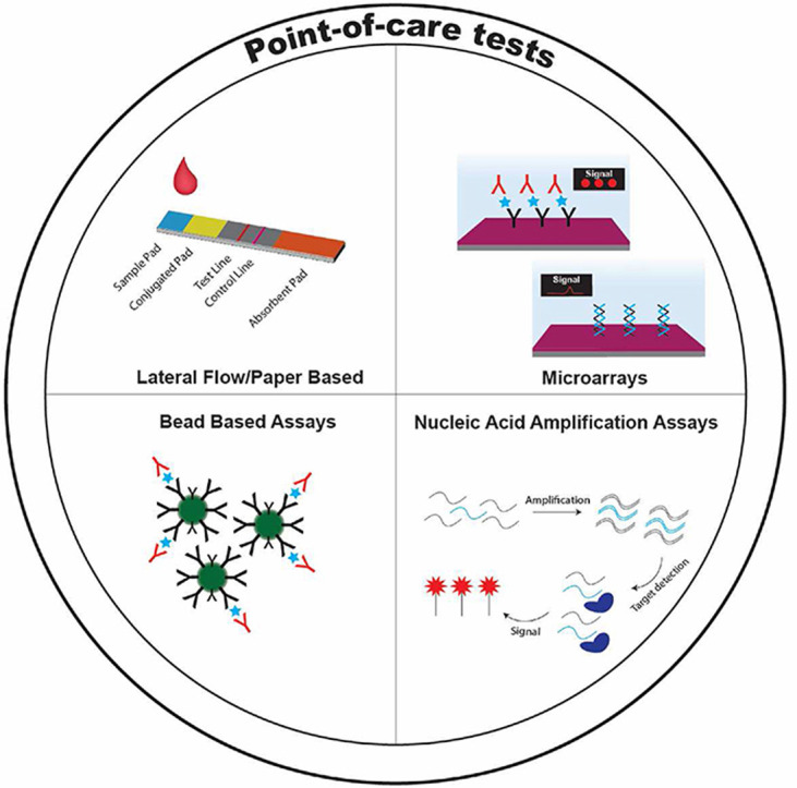
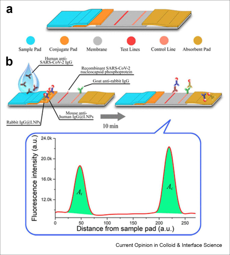
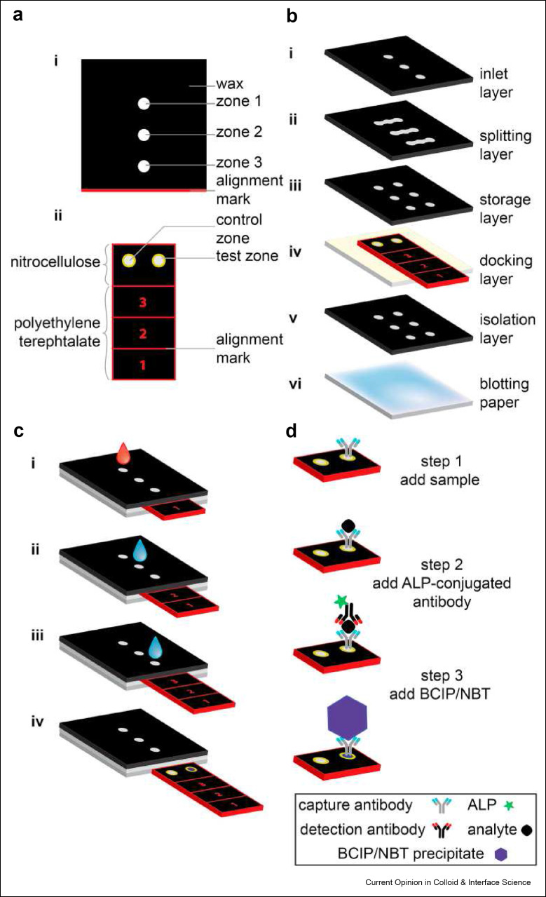
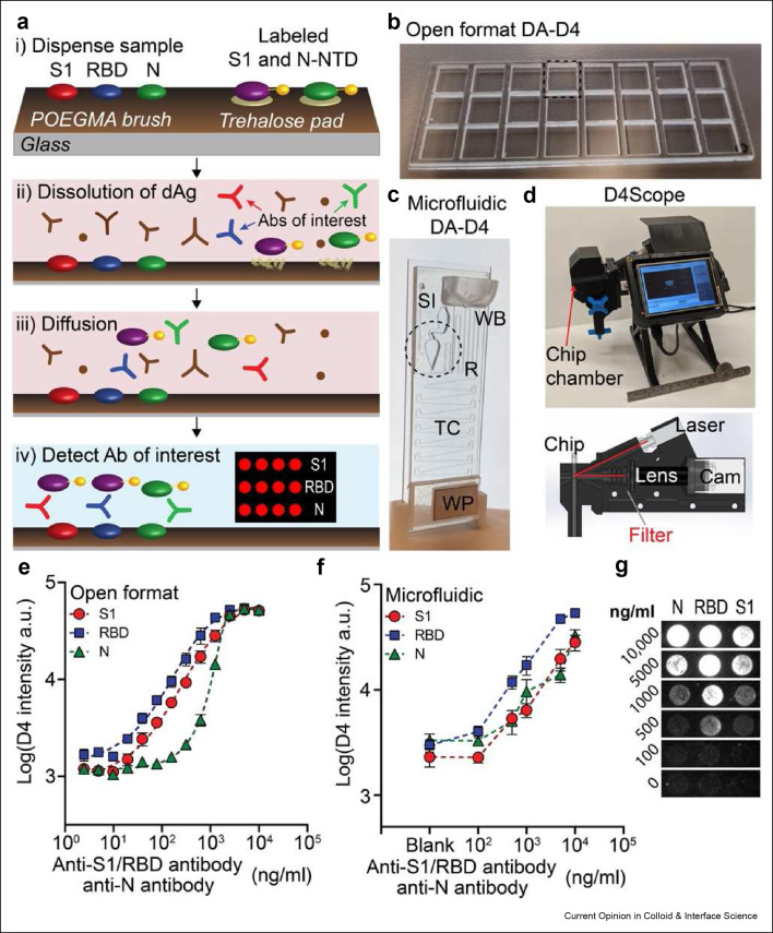
The ongoing Coronavirus disease 2019 (COVID-19) pandemic illustrates the need for sensitive and reliable tools to diagnose and monitor diseases. Traditional diagnostic approaches rely on centralized laboratory tests that result in long wait times to results and reduce the number of tests that can be given. Point-of-care tests (POCTs) are a group of technologies that miniaturize clinical assays into portable form factors that can be run both in clinical areas --in place of traditional tests-- and outside of traditional clinical settings --to enable new testing paradigms. Hallmark examples of POCTs are the pregnancy test lateral flow assay and the blood glucose meter. Other uses for POCTs include diagnostic assays for diseases like COVID-19, HIV, and malaria but despite some successes, there are still unsolved challenges for fully translating these lower cost and more versatile solutions. To overcome these challenges, researchers have exploited innovations in colloid and interface science to develop various designs of POCTs for clinical applications. Herein, we provide a review of recent advancements in lateral flow assays, other paper based POCTs, protein microarray assays, microbead flow assays, and nucleic acid amplification assays. Features that are desirable to integrate into future POCTs, including simplified sample collection, end-to-end connectivity, and machine learning, are also discussed in this review.

Keywords: COVID-19; Point-of-care tests; biosensors; in vitro diagnostics; infectious disease.


Conflict of interest statement

☒ The authors declare the following financial interests/personal relationships which may be considered as potential competing interests: Ashutosh Chilkoti reports financial support was provided by National Institutes of Health. Ashutosh Chilkoti has patent #WO/2020/223713 issued to Duke University. Ashutosh Chilkoti has patent #PCT/US2021/046833 issued to Duke University. Ashutosh Chilkoti has patent #63/429,316 pending to Duke University. David Kinnamon has patent #PCT/US2021/046833 issued to Duke University. Jacob Heggestad has patent #PCT/US2021/046833 issued to Duke University. David Kinnamon has patent #63/429,316 pending to Duke University. Jacob Heggestad has patent #63/429,316 pending to Duke University. Immucor Inc. has acquired the rights to the D4 assay on POEGMA brushes for in vitro diagnostics from Sentilus Inc. (cofounded by A.C. and others). D.T.B declares no competing interests.

Figures

Image 1
Graphical abstract
Figure 1
Figure 1
A) Design of a LFA to detect anti-SARS-CoV-2 antibodies. B) Simulated assay using lanthanide doped up-converting phosphor nanoparticles as the fluorescent reporter. Reproduced with permission from ACS:.https://pubs.acs.org/doi/10.1021/acs.analchem.0c00784. Please note that further permissions related to the use of this material should be directed to the ACS. Copyright (2020), (ACS Analytical Chemistry).
Figure 2
Figure 2
Design schematic of the sliding strip microfluidic device by Verma et al. A) Top view of the device. B) Exploded view of each component of the sliding strip microfluidic. C) Stepwise operation of the sliding strip device. Step 1, addition of sample and water wash. Step 2, water addition to dissolve stored detection antibodies and buffer. Step 3, addition of more water to dissolve substrates and a buffer needed to complete the reaction and generate a colorimetric signal. Step 4, removal of sliding strip for results readout. Results can be captured visually or by using a desktop scanner. D) Secondary outline of steps with an emphasis on the molecular interactions happening at each stage of the sliding strips checkpoints. Reused with permission under creative copyright license CC-BY 4.0 2018 (Elsevier).
Figure 3
Figure 3
A) Design of the D4 assay for anti-SARS-CoV-2 antibodies. B) POCT format at the open format D4 assay. C) Image of the POCT microfluidic D4 cassette. D) Image of the D4Scope. E) Quantitative performance of the open format D4 assay for anti-SARS-CoV-2 antibodies. F) Quantitative performance of the microfluidic D4 assay for anti-SARS-CoV-2 antibodies. G) Spot image representation for assay intensity at each dose point. Reused with permission under creative copyright license CC-BY-NC 4.0 2021 (AAAS).
Figure 5
Figure 5
Schematic of SHERLOCK. A) RNA is extracted from clinical samples and amplified by either reverse transcriptase RPA or standard RPA before being placed into a reaction mixture that contains T7 RNA polymerase, Cas13, target specific crRNA, and a fluorescent RNA reporter, which produces a fluorescence signal. B) Fluorescence read-out from SHERLOCK of patient samples acquired during the 2015-2016 Zika pandemic. C) Comparison of SHERLOCK (left and middle columns) with Altona RT-PCR results (right column). D) Fluorescence output from SHERLOCK for DENV positive clinical samples and controls. Reused with permission under creative copyright license CC-BY 4.0 2018 (AAAS).
Figure 7
Figure 7
Schematic of SENSR, a one-pot isothermal reaction cascade for the rapid detection of RNA. The reaction is composed of four main components: a set of probes, SplintR ligase, T7 RNA polymerase and a fluorogenic dye. In the presence of target RNA, hybridization, ligation, transcription and aptamer–dye binding reactions occur sequentially in a single reaction tube at a constant temperature and yields a fluorescence signal. Reused with permission under creative copyright license CC-BY 4.0 2020 (Springer Nature).
Figure 6
Figure 6
SIMPLE microfluidic device. A) User places whole blood samples onto the chip and sample processing proceeds automatically. B) DNA is amplified via a reusable heat pack at a constant temperature. C) Dye loaded chip for visual presentation. Red dye illustrates the microfluidic channels, blue dye illustrates the primary vacuum and battery system, green dye shows the auxiliary vacuum. D) Digital micropatterning for red blood cell separation (left) and an amplification initiator stencil for RPA (right) scale bars are 1 mm (yellow) and 100 μm (black). E) Profile view of whole blood separation within the microfluidic region. F) On-board vacuum to provide a portable power supply. Reused with permission under creative copyright license CC-BY 4.0 2017 (AAAS).
Figure 4
Figure 4
A) Comparison of graphene FET in planar (left) and bent and crumpled (right) orientations. B) Fabrication process for graphene shrinking and probe DNA immobilization (black) and target DNA (red) hybridization. C) SEM images of graphene in the bent and crumpled state, left scale bar is 5 μM, right scale bar is 500 nm. D) Raman spectrum of graphene on the polystyrene substrate. E) Charge transfer of the crumpled graphene highlighting the shift in the electron transfer at the Dirac point. F) Dirac point shift as a function of pH. Reused with permission under creative copyright license CC-BY 4.0 2020 (Springer Nature).
Figure 8
Figure 8
SiMoA assay overview. A) Bead labeled with capture antibodies capture a target biomarker —the analyte— which is then bound by a second antibody that is conjugated to an enzymatic reporter. B) Beads are then loaded into femtoliter wells for fluorescence detection. C) SEM of femtoliter wells after bead loading. D) Fluorescence image of the beads in wells. Reused with permission under creative copyright license CC-BY 4.0 2010 (Springer Nature).
Figure 9
Figure 9
TAP microneedle blood collection device. Left: underside of the device showing the flow channel and heparinized reservoir. Right: 3D transparent device view of the blood flow channel, heparinized reservoir, and fill completion indicator. Reused with permission under creative copyright license CC-BY 4.0 2018 (Springer Nature).
Figure 10
Figure 10
Artificial intelligence enhanced POCT For HPV detection. A) Assay procedure including obtaining cells from cervical brushing (far left), hybridization to PS and silica beads (middle left), diffraction imaging of the dimer hybrids (middle right), and deep learning analysis algorithm (far right). B) Photograph of AIM-HPV device. C) 3-D rendering of AIM-HPV device showing the display, switch, LED, diffuser, pinhole, CMOS imager, power port, and microUSB port. D) Diffraction images showing PS bead monomers (blue arrows), silica bead monomers (orange arrows), and PS-silica dimer (red arrow). Reused with permission under creative copyright license CC-BY 4.0 2019 (Ivyspring International).

Similar articles

Cited by

References

    1. Kozel T.R., Burnham-Marusich A.R. Point-of-Care Testing for Infectious Diseases: Past, Present, and Future. Journal of Clinical Microbiology. 2017;55(8):2313. doi: 10.1128/JCM.00476-17. - DOI - PMC - PubMed
    1. Yang S., Rothman R.E. PCR-based diagnostics for infectious diseases: uses, limitations, and future applications in acute-care settings. The Lancet Infectious Diseases. 2004;4(6):337–348. doi: 10.1016/S1473-3099(04)01044-8. 2004/06/01/ - DOI - PMC - PubMed
    1. Wang S., Lifson M.A., Inci F., Liang L.G., Sheng Y.F., Demirci U. Advances in addressing technical challenges of point-of-care diagnostics in resource-limited settings. Expert Rev Mol Diagn. 2016;16(4):449–459. doi: 10.1586/14737159.2016.1142877. - DOI - PMC - PubMed
    1. Sosnik A., Amiji M. Nanotechnology solutions for infectious diseases in developing nations. Advanced Drug Delivery Reviews. 2010;62(4):375–377. doi: 10.1016/j.addr.2009.11.010. - DOI - PubMed
    1. Wang C., Horby P.W., Hayden F.G., Gao G.F. A novel coronavirus outbreak of global health concern. The Lancet. 2020;395(10223):470–473. doi: 10.1016/S0140-6736(20)30185-9. - DOI - PMC - PubMed

LinkOut - more resources