. 2023 Jun 8;8(24):21793-21801.
doi: 10.1021/acsomega.3c01374. eCollection 2023 Jun 20.

Functionalized Magnetic Nanoparticles for NIR-Induced Photothermal Therapy of Potential Application in Cervical Cancer

Affiliations

Functionalized Magnetic Nanoparticles for NIR-Induced Photothermal Therapy of Potential Application in Cervical Cancer

Linxue Zhang et al. ACS Omega. .

Abstract

Photothermal therapy (PTT) holds great promise for cancer treatment with its effective ablation of solid tumors. As the essential core point, photothermal agents (PTAs) with excellent photothermal properties and good biocompatibility could help to fulfill highly efficient PTT. Herein, a novel type of nanoplatform Fe3O4@PDA/ICG (FPI) nanoparticle (NP) was designed and synthesized, which was composed of magnetic Fe3O4 and near-infrared excitable indocyanine green via encapsulation of polydopamine. The FPI NPs showed spherical structures in shape with uniform distribution and good chemical stability. Under 793 nm laser irradiation, FPI NPs could generate hyperthermia of 54.1 °C and photothermal conversion efficiency of 35.21%. The low cytotoxicity of FPI NPs was further evaluated and confirmed on HeLa cells with a high survival rate (90%). Moreover, under laser irradiation (793 nm), FPI NPs showed effective photothermal therapeutic characteristics for HeLa cells. Therefore, FPI NPs, as one of the promising PTAs, have great potential in the field of PTT for tumor treatment.


Conflict of interest statement

The authors declare no competing financial interest.

Figures

Figure 1
Figure 1
Morphology image of (A), (B) Fe3O4 structure, (C) XRD patterns of Fe3O4 NPs, morphology image of (D), (E) FPI structures. (F) Zeta potential of ICG, PDA, Fe3O4, FP, and FPI NPs.
Scheme 1
Scheme 1. NIR-Emissive FPI NPs for Target-Assisted PTT for Cervical Cancer
Figure 2
Figure 2
Photothermal performance of samples under 793 nm laser irradiation at a power density of 0.33 W/cm2: (A) Temperature curves of ICG, Fe3O4, and FPI NPs in the aqueous dispersion (100 μg/mL) at various time points, respectively; (B) images of ICG, PDA, Fe3O4, and FPI NPs in the aqueous dispersion (from left to right, respectively); (C) temperature curves of FPI NP dispersions with various concentrations (20, 40, 60, 80, and 100 μg/mL); (D) NIR-thermal images for FPI NPs of different concentrations (20, 40, 60, 80, and 100 μg/mL).
Figure 3
Figure 3
(A) Photothermal stability of FPI NPs (100 μg/mL) during five cycles with/without laser (793 nm, 0.33 W/cm2). (B) Temperature evaluation of FPI NPs (100 μg/mL) with/without laser (793 nm, 0.33 W/cm2), and linear time versus -Lnθ data of FPI NPs (during the cooling period). (C) Images of FPI NPs before (left)/after (right) laser (100 μg/mL, 793 nm, and 0.33 W/cm2). (D) Temperature evaluation of free Fe3O4 (100 μg/mL) with/without laser (793 nm, 0.33 W/cm2), and linear time versus -Lnθ data of free Fe3O4 (during the cooling period).
Figure 4
Figure 4
(A) Fluorescence image of Fe3O4 and FPI NPs (concentration of 100 mg/mL) under the CLSM (λem = 800–1000 nm, λex = 640 nm). (B) HeLa cell cytotoxicity viability by CCK-8 of HeLa cells after incubation with Fe3O4 and FPI NPs at different concentrations (0, 20, 40, 80, and 100 μg/mL); (C) HeLa cell viability of Fe3O4 and FPI NPs at different concentrations (0, 20, 40, 80, and 100 μg/mL), assessed on treated cells that were laser-irradiated at 0.33 W/cm2 for 10 min. (D) Flow cytometry in the case of Control, Fe3O4 (40 μg/mL), and FPI NPs (40 μg/mL). Confocal microscopy of cells in bright field (E) HeLa cell (control), (F) after HeLa cell treatment with FPI NPs for 4 h.
Figure 5
Figure 5
CLSM images of the samples (40 μg/mL) in HeLa cells treated with different conditions (DMEM, Fe3O4, FPI, Fe3O4 and laser, and FPI and laser) (scale bar = 500 μm).
Figure 6
Figure 6
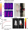
Under 793 nm NIR light irradiation (A) infrared thermal images of the mice treated with PBS and FPI NPs. (B) Temperature change curve of the subcutaneous injection site. (C) NIR fluorescence images of mice through the subcutaneous injection with PBS and FPI NPs at 2 h.

Similar articles

Cited by

References

    1. Alifu N.; Ma R.; Zhu L. J.; Du Z.; Chen S.; Yan T.; Alimu G.; Zhang L. X.; Zhang X. L. A Novel TMTP1-modified theranostic nanoplatform for targeted in vivo NIR-II fluorescence imaging-guided chemotherapy of cervical cancer. J. Mater. Chem. B 2022, 10, 506–517. 10.1039/D1TB02481G. - DOI - PubMed
    1. Zhu L. J.; Chen J. J.; Yan T.; Alimu G.; Zhang X. L.; Chen S.; Aimaiti M.; Ma R.; Alifu N. Near-infrared emissive polymer-coated IR-820 nanoparticlesassisted photothermal therapy for cervical cancer cells. J. Biophotonics. 2021, 14, e20210011710.1002/jbio.202100117. - DOI - PubMed
    1. Shrestha A. D.; Neupane D.; Vedsted P.; Kallestrup P. Cervical cancer prevalence, incidence and mortality in low and middle income countries: a systematic review. Asian Pac. J. Cancer Prev. 2018, 19, 319–324. 10.22034/APJCP.2018.19.2.319. - DOI - PMC - PubMed
    1. Asami Y.; Yutaka U.; Mamoru K.; Yusuke T.; Sayaka I.; Shinya M.; Eiji K.; Toshitaka M.; Isao M.; Keisuke F.; Yuri I.; Tomio N.; Tadashi K. Epidemiologic and clinical analysis of cervical cancer using data from the population-based Osaka cancer registry. Cancer Res. 2019, 79, 1252–1259. 10.1158/0008-5472.CAN-18-3109. - DOI - PubMed
    1. Liu S.; Pan X.; Liu H. Two-dimensional nanomaterials for photothermal therapy. Angew. Chem. Int. Ed. 2020, 132, 5943–5953. 10.1002/ange.201911477. - DOI - PubMed