. 2023 Jun 22;12(6):e1455.
doi: 10.1002/cti2.1455. eCollection 2023.

A novel dual NLRP1 and NLRP3 inflammasome inhibitor for the treatment of inflammatory diseases

Affiliations

A novel dual NLRP1 and NLRP3 inflammasome inhibitor for the treatment of inflammatory diseases

Callum Ah Docherty et al. Clin Transl Immunology. .

Abstract

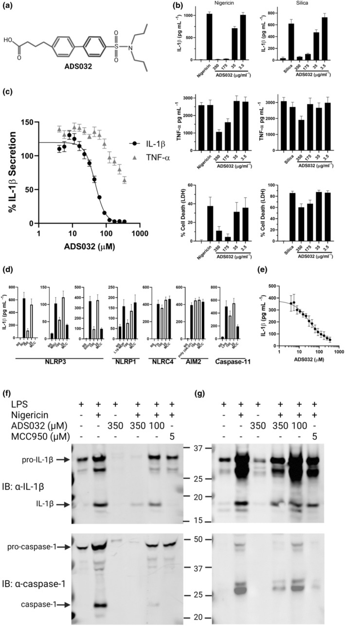
Objectives: Inflammasomes induce maturation of the inflammatory cytokines IL-1β and IL-18, whose activity is associated with the pathophysiology of a wide range of infectious and inflammatory diseases. As validated therapeutic targets for the treatment of acute and chronic inflammatory diseases, there has been intense interest in developing small-molecule inhibitors to target inflammasome activity and reduce disease-associated inflammatory burden.

Methods: We examined the therapeutic potential of a novel small-molecule inhibitor, and associated derivatives, termed ADS032 to target and reduce inflammasome-mediated inflammation in vivo. In vitro, we characterised ADS032 function, target engagement and specificity.

Results: We describe ADS032 as the first dual NLRP1 and NLRP3 inhibitor. ADS032 is a rapid, reversible and stable inflammasome inhibitor that directly binds both NLRP1 and NLRP3, reducing secretion and maturation of IL-1β in human-derived macrophages and bronchial epithelial cells in response to the activation of NLPR1 and NLRP3. ADS032 also reduced NLRP3-induced ASC speck formation, indicative of targeting inflammasome formation. In vivo, ADS032 reduced IL-1β and TNF-α levels in the serum of mice challenged i.p. with LPS and reduced pulmonary inflammation in an acute model of lung silicosis. Critically, ADS032 protected mice from lethal influenza A virus challenge, displayed increased survival and reduced pulmonary inflammation.

Conclusion: ADS032 is the first described dual inflammasome inhibitor and a potential therapeutic to treat both NLRP1- and NLRP3-associated inflammatory diseases and also constitutes a novel tool that allows examination of the role of NLRP1 in human disease.

Keywords: NLRP1; NLRP3; drug targets; inflammasome; inflammation; pulmonary inflammation.


Conflict of interest statement

RED and MAN receive consultancy fees from Adiso Therapeutics Inc. RF, DLF, CKM and AM are employees of Adiso Therapeutics Inc. AJF and AGR are supported by an Adiso Therapeutics Inc research grant to the University of Edinburgh Centre for Inflammation Research. RED, MAN, RF and CKM have patent ownership, while CKM and AM retain stock ownership in Adiso Therapeutics.

Figures

Figure 1
Figure 1
ADS032 is a novel NLRP3 inhibitor. (a) ADS032 structure. (b) Immortalised bone marrow‐derived macrophages (iBMDMs) were seeded at 4 × 105 mL−1 prior to priming with LPS (100 ng mL−1) for 3 h. Media were removed 60 min prior to challenge and replaced with serum‐free media containing ADS032 (3.5–350 μm) as indicated, or DMSO control (0.8% v/v). Macrophages were stimulated with either nigericin (6 μm) or silica (250 μg mL−1) as indicated for 120 or 360 min respectively. Cellular supernatants were analysed for secreted IL‐1β, TNF‐α by ELISA or cell death by LDH assay as indicated. (c) iBMDMs were treated with a range of ADS032 concentrations for 60 min in serum‐free media prior to challenge with nigericin for 120 min. The results shown are representative of three independent experiments carried out in triplicate and presented as the mean ± SEM. Non‐linear regression analysis was performed, and the curve of the log [M] ADS032 versus the normalised response (variable slope) is presented. (d) LPS (3 h;100 ng mL−1)‐primed iBMDMs were pre‐treated with ADS032 (20 and 100 μg mL−1) or MCC950 (MCC: 5 μg mL−1) in serum‐free media for 60 min and treated with NLRP3 (nigericin, 6 μm, 120 min; monosodium urate (MSU), 250 μg mL−1, 6 h; and silica, 250 μg mL−1; 6 h), NLRP1 (L18‐MDP, 100 μg mL−1, 16 h), non‐canonical NLRP3 (transfected LPS Escherichia coli serotype 0111 B4, 2.0 μg mL−1, 16 h), AIM2 (poly dA:dT, 1 μg mL−1, 8 h) and NLRC4 (flagellin, 20 μg mL−1, 16 h) agonists as indicated. Secreted IL‐1β was assayed by ELISA and the results shown are representative of three independent experiments carried out in triplicate and presented as the mean ± SEM. (e) IC50 value of L18‐MDP (NLRP1) activity was determined as normalised IL‐1β secretion in ADS032‐treated iBMDMs. (f) BMDMs generated from wild‐type mice, and (g) PMA‐differentiated THP‐1 macrophages were primed with LPS for 3 h, treated with ADS032 (100 and 300 μm) or MCC950 (5 μm) where indicated for 60 min, then subsequently challenged with nigericin for 180 min. Cultured supernatants were assayed for secreted IL‐1β and caspase‐1 following 4–12% SDS‐PAGE, transfer to PVDF membrane and immunoblotting (IB) with indicated antibodies. The results shown are representative of three independent experiments.
Figure 2
Figure 2
ADS032 directly binds to NLRP1 and NLRP3. (a, b) ADS165 and ADS167 structure. (c) Recombinant NLRP3 (2 μg) or (e) NLRP1 (2 μg) was co‐incubated where indicated with ADS167 or MCC950 for 20 min and then irradiated with UV 365 nm for a further 20 min. ADS165 was then co‐incubated for 20 min and UV treated for 20 min to cross‐link‐associated compound and protein. Protein was separated by SDS‐PAGE and immunoblotted (IB) with anti‐PEG to visualise ADS165‐linked protein. (d) iBMDM cells stably expressing NLRP3‐Flag were treated for 30 min with ADS165, ADS167 (1 mm) or MCC950 (50 μm) for 30 min, and irradiated with UV 365 nm for 30 min. Cells were lysed with buffer and immunoprecipitation (IP) of NLRP3 was performed with α‐NLRP3 (D4D8T) antibody. Levels of precipitated (IP) and total cellular lysate (Input) NLRP3 were determined by immunoblotting with α‐NLRP3 (Cryo‐2) antibody. (f) NLRP1‐Flag was expressed in HEK293T cells (1 × 106) for 20 h and treated with ADS165 (1 mm) for 30 min where indicated, followed by UV 365 nm for a further 30 min. Cells were lysed with lysis buffer and NLRP1 immunoprecipitated with α‐Flag (M2) antibody. Proteins were separated by SDS‐PAGE and ADS165‐linked NLRP1 identified by immunoblot with α‐PEG, while total precipitated NLRP1 was visualised by immunoblot with α‐NLRP1 and total cell lysate by α‐Flag. The results shown are representative of three independent experiments.
Figure 3
Figure 3
ADS032 dose dependently reduces NLPR3‐induced ASC oligomerisation. ASC‐tagged‐Cerulean (pseudo‐coloured Red) cells were treated or not with ADS032 (20–350 μm) or MCC950 (MCC: 5 μm) for 60 min and then challenged with either (a) nigericin (6 μm, 120 min) or (b) silica (250 μg mL−1, 5 h) before fixation. Cells were stained with Hoechst 33342 to identify cell nuclei (Blue). Images are flattened maximum‐intensity projections of three‐dimensional deconvolved z stacks. Specks were observed with confocal microscopy (magnification, ×630) and (c) the percentage of specks per field of view (nine fields per treatment group) were counted and compared to agonist‐treated cells. NS, not stimulated. Data presented are representative of three independent experiments.
Figure 4
Figure 4
ADS032 effectively inhibits NLRP1 and NLRP3 inflammasome activity in human macrophages and bronchial epithelial cells. PMA‐differentiated THP‐1 cells primed with LPS (100 ng mL−1) for 3 h, pre‐treated or not with ADS032 (35–350 μm) for 60 min and then challenged with (a) NLRP3 agonists nigericin (6 μm) or silica (250 μg mL−1) for 120 or 360 min, respectively, or (c) NLPR1 agonists L18‐MDP (100 μg mL−1) or transfected poly I:C (200 ng mL−1) for 16 or 8 h respectively. (b) Human monocyte‐derived macrophages were treated for 3 h with 50 pg mL−1 LPS, treated with ADS032 (20–350 μm) or MCC950 (MCC; 5 μm) in serum‐free media for 60 min, then challenged with nigericin (6 μm) for a further 120 min. Bronchial epithelial cells obtained from normal patients were treated with ADS032 as indicated for 60 min and (d) treated with poly I:C (200 ng mL−1) for a further 8 h or (e) talabostat (2.5 μm; 24 h) and anisomycin (2.0 μm; 8 h). Cultured supernatants were assayed for secreted IL‐1β by ELISA. The results shown are the pooled data of three independent experiments carried out in triplicate and presented as the mean ± SEM.
Figure 5
Figure 5
ADS032 is a rapidly acting, long‐lasting, reversible NLRP3 inhibitor. LPS (100 ng mL−1)‐primed iBMDM cells (2 × 105 mL−1) were (a) treated with ADS032 (100 μm) prior (i.e. −10 to −60 min), simultaneously (i.e. 0 min) or post (i.e. 10 or 30 min) nigericin challenge, as indicated. (b) ADS032 (350 μm) was administered to primed iBMDMs for between 1 and 8 h prior to nigericin challenge for a further 120 min. (c) Primed iBMDMs were treated with ADS032 (20–350 μm) in the final 60 min of LPS priming and subsequently stimulated with nigericin for a further 2 h. ADS032 was either left on cells (No Washout) or removed (Washout) for 1 min prior to nigericin challenge. Secreted IL‐1β was assessed by ELISA and the data are representative of three independent experiments conducted in triplicate and presented as the mean ± SEM. Non‐linear regression analysis was performed (c), and the curve of the log [M] ADS032 versus the normalised response (variable slope) is presented. The results shown are representative of three independent experiments carried out in triplicate and shown as the mean ± SEM.
Figure 6
Figure 6
ADS032 reduced systemic inflammation in vivo. Serum levels of C57Bl/6 wild‐type mice (n = 8; 4 male/4 female per group) pre‐treated with ADS032 (200 mg kg−1) or vehicle control (methylcellulose) for 60 min prior to LPS challenge (10 mg kg−1, i.p.) for 120 min demonstrated reduced levels of IL‐1β, TNF‐α and IFN‐γ, but not IL‐10, as determined by ELISA or cytokine bead array. Data are presented as representative of two independent experiments. *P < 0.05, **P < 0.01 and ***P < 0.001, ns = not significant by the Mann–Whitney test.
Figure 7
Figure 7
ADS032 reduces lung inflammation in an acute model of silicosis. Wild‐type C57Bl/6 mice (n = 6 per treatment group) received PBS alone, silica (50 mg kg−1), silica (50 mg kg−1) mixed with ADS132 (40 mg kg−1) in PBS or ADS0132 alone (40 mg kg−1) via intranasal inoculation. Twenty‐four hours later, mice were killed and bronchial alveolar lavage fluid was harvested. Proinflammatory cytokines (a) IL‐1β, (b) TNF‐α, (c) MCP‐1 and (d) IL‐6 were examined by ELISA or cytokine bead array. Total numbers of (e) leukocytes in BALF were determined by viable cell counts, (f) Ly6G+ neutrophils, (g) Ly6C+ inflammatory macrophages and (h) CD11c+ I‐Abhigh dendritic cells were determined by flow cytometry. The data presented are representative of two independent experiments and are the means ± SEM. *P < 0.05, ***P < 0.001, ns = not significant. One‐way ANOVA (multiple comparisons).
Figure 8
Figure 8
ADS032 therapeutically protects mice from severe IAV disease and reduces pulmonary inflammation. Groups of male and female C57Bl/6 mice (n = 8 per group) were intranasally challenged with HKx31 (105 PFU) IAV. Mice were treated intranasally with PBS (IAV) or ADS032 (20 mg kg−1) from either day 1 or day 3 post‐infection and every 48 h thereafter. Uninfected mice (n = 4 per group) treated with PBS or ADS032 were included for comparison. (a) Mice were weighed daily and results were expressed as the mean ± SEM. (b) Survival curves are shown. ***P < 0.001. Mantel–Cox log‐rank test. Groups of mice, as described above, were also challenged with HKx31 (105 PFU) and treated with ADS032 (20 mg kg−1) every 48 h from day 1. On day 4 post‐infection, mice were killed. Proinflammatory cytokines (c) IL‐1β, (d) IL‐6, (e) TNF‐α and (f) MCP‐1 were determined by ELISA or cytokine bead array in BALF. Total numbers of (g) leukocytes in BALF were determined by viable cell counts, and (h) Ly6G+ neutrophils, (i) total CD11c+ I‐Ablow macrophages and (j) CD11c+ I‐Abhigh dendritic cells were determined by flow cytometry. The results shown are representative of two independent experiments and presented are the means ± SEM from eight mice in IAV/IAV‐ADS032 groups, and four mice in PBS/ADS032 groups. *P < 0.05, **P < 0.01 and ***P < 0.001. One‐way ANOVA.

Similar articles

Cited by

References

    1. Pandey A, Shen C, Feng S, Man SM. Cell biology of inflammasome activation. Trends Cell Biol 2021; 31: 924–939. - PubMed
    1. Schroder K, Tschopp J. The inflammasomes. Cell 2010; 140: 821–832. - PubMed
    1. Broz P, Dixit VM. Inflammasomes: mechanism of assembly, regulation and signalling. Nat Rev Immunol 2016; 16: 407–420. - PubMed
    1. Ross C, Chan AH, von Pein JB, Maddugoda MP, Boucher D, Schroder K. Inflammatory caspases: toward a unified model for caspase activation by inflammasomes. Annu Rev Immunol 2022; 40: 249–269. - PubMed
    1. Broz P, Pelegrin P, Shao F. The gasdermin, a protein family executing cell death and inflammation. Nat Rev Immunol 2020; 20: 143–157. - PubMed