Case Reports
. 2023 Jun 15:2023:7005565.
doi: 10.1155/2023/7005565. eCollection 2023.

Ectopic Cecal Varices as a Cause of Lower Gastrointestinal Bleeding

Affiliations
Case Reports

Ectopic Cecal Varices as a Cause of Lower Gastrointestinal Bleeding

Abdulrahman Qatomah et al. Case Rep Gastrointest Med. .

Abstract

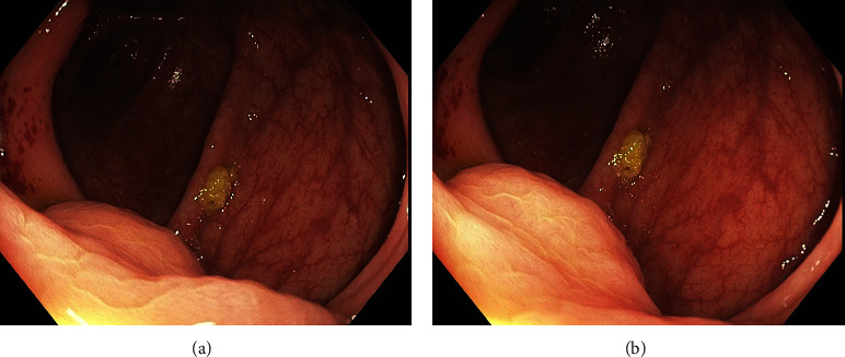
Ectopic varices account for 1%-5% of all variceal bleeding episodes in patients with portal hypertension. They can be found at any part of gastrointestinal tract including the small intestines, colon, or rectum. We report a case of a 59-year-old man who presented with bleeding per rectum 2 days after a routine colonoscopy, in which 2 lesions were biopsied. Gastroscopy was negative for bleeding, and he was not stable enough to undergo colonoscopy. CT angiography showed a large portosystemic shunt with multiple collaterals in the right lower quadrant. These findings were clues for a diagnosis of ectopic cecal varices.


Conflict of interest statement

The authors declare that they have no conflicts of interest.

Figures

Figure 1
Figure 1
(a, b) Endoscopic image with semi-circumferential lesion just above the ileocecal valve without the typical appearance of varices.
Figure 2
Figure 2
Abdomen CT ((a) axial and (b) coronal) showing portosystemic shunt with multiple collaterals in the right lower quadrant, in between the superior mesenteric vein and inferior vena cava (red arrows).
Figure 3
Figure 3
(a, b) Endoscopic image showing the cecum after sclerotherapy (thrombin) with no evidence of active bleeding.

References

    1. Helmy A., Al Kahtani K., Al Fadda M. Updates in the pathogenesis, diagnosis and management of ectopic varices. Hepatology International . 2008;2(3):322–334. doi: 10.1007/s12072-008-9074-1. - DOI - PMC - PubMed
    1. Sarin S. K., Kumar C. K. N. Ectopic varices. Clinical Liver Disease . 2012;1(5):168–172. doi: 10.1002/cld.95. - DOI - PMC - PubMed
    1. Schreiner P., Puippe G., Gubler C. An unusual, but potentially life-threatening lesion in the cecum. Gastroenterology . 2018;155(2):e9–e10. doi: 10.1053/j.gastro.2017.12.036. - DOI - PubMed
    1. Haddad J. D., Lacey B. W. Isolated non-hemorrhagic cecal varices. Gastroenterology Report . 2014;2(4):316–319. doi: 10.1093/gastro/gou027. - DOI - PMC - PubMed
    1. Kudaravalli P., Saleem S. A., Pendela V. S., Manocha D. A therapeutic approach to cecal varices. ACG Case Reports Journal . 2020;7(2) doi: 10.14309/crj.0000000000000315.e00315 - DOI - PMC - PubMed

Publication types

LinkOut - more resources