Case Reports
. 2023;32(3):161-167.
doi: 10.1297/cpe.2022-0071. Epub 2023 Apr 14.

A case of hyperphosphatemic familial tumoral calcinosis due to maternal uniparental disomy of a GALNT3 variant

Affiliations
Case Reports

A case of hyperphosphatemic familial tumoral calcinosis due to maternal uniparental disomy of a GALNT3 variant

Naoko Nishimura-Kinoshita et al. Clin Pediatr Endocrinol. 2023.

Abstract

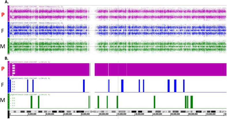
Hyperphosphatemic familial tumoral calcinosis (HFTC) is a rare, inherited autosomal recessive disorder caused by fibroblast growth factor-23 (FGF23), N-acetylgalactosaminyltransferase 3 (GALNT3), or Klotho (KL) gene variants. Here, we report the case of a Japanese boy who presented with a mass in his left elbow at the age of three. Laboratory test results of the patient revealed normocalcemia (10.3 mg/dL) and hyperphosphatemia (8.7 mg/dL); however, despite hyperphosphatemia, serum intact FGF23 level was low, renal tubular reabsorption of phosphate (TRP) level was inappropriately increased, and 1,25-dihydroxyvitamin D3 (1,25(OH)2D3) level was inappropriately normal. Genetic analysis revealed maternal uniparental disomy (UPD) of chromosome 2, which included a novel GALNT3 variant (c.1780-1G>C). Reverse transcription-polymerase chain reaction (RT-PCR) analysis of GALNT3 mRNA confirmed that this variant resulted in the destruction of exon 11. We resected the mass when the patient was five years old, owing to its gradual enlargement. No relapse or new pathological lesions were observed four years after tumor resection. This is the first case report of a Japanese patient with HFTC associated with a novel GALNT3 variant, as well as the first case of HFTC caused by maternal UPD of chromosome 2 that includes the GALNT3 variant.

Keywords: N-acetylgalactosaminyltransferase 3 (GALNT3); hyperostosis–hyperphosphatemia syndrome; hyperphosphatemic familial tumoral calcinosis; tumoral calcinosis; uniparental disomy.


Figures

Fig. 1.
Fig. 1.
Ectopic multilocular calcification in the patient. A: X-ray and B: coronal T2-weighted magnetic resonance images of the left elbow at the first visit (3 yr and 7 mo old). The tumor enlarged and caused self-destruction of the skin at 5 yr (y) and 1 mo (m) of age.
Fig. 2.
Fig. 2.
GALNT3 variant identified in our patient. A: The DNA sequence analysis of our proband revealed a splice site-affecting homozygous variant in intron 10 (c.1780-1G>C). B: Schematic design of GALNT3 mRNAs and PCR products. We designed a primer set to amplify each exon as mapped below the GALNT3 mRNA model. C: The asymptomatic mother and sister of the proband had the same variant (heterozygous), whereas his father did not. D: PCR analysis confirmed the deletion of exon 11 in the GALNT3 mRNA of the patient. As described in panel C, the primer sets 11-1 and 11-2 were designed to amplify exons 10 and 11. The PCR products amplified from the cDNA of healthy donors were of the expected size, whereas these bands were absent in the cDNA of the patient (D). WBC, white blood cell.
Fig. 3.
Fig. 3.
Genome-wide SNP array indicating maternal isodisomy of chromosome 2. A: Differences in the alleles of genotyped SNPs on chromosome 2 in the proband (P), his father (F), and his mother (M) are displayed. B: Loss of heterozygosity (LOH) on chromosome 2. LOH represents the loss of allelic differences, and the LOH state is either 0, representing no LOH or 1, representing LOH. Allele segregation in the proband comprised homozygotes and LOH; however, parental (F and M) allele segregation exhibited a heterozygous pattern on chromosome 2. The proband had 7,673 homozygous SNPs from the mother and six homozygous SNPs from the father.

References

    1. Ramnitz MS, Gourh P, Goldbach-Mansky R, Wodajo F, Ichikawa S, Econs MJ, et al. . Phenotypic and genotypic characterization and treatment of a cohort with familial tumoral calcinosis/hyperostosis-hyperphosphatemia syndrome. J Bone Miner Res 2016;31: 1845–54. doi: 10.1002/jbmr.2870 - DOI - PMC - PubMed
    1. Boyce AM, Lee AE, Roszko KL, Gafni RI. Hyperphosphatemic tumoral calcinosis: pathogenesis, clinical presentation, and challenges in management. Front Endocrinol (Lausanne) 2020;11: 293. doi: 10.3389/fendo.2020.00293 - DOI - PMC - PubMed
    1. Engel E. A fascination with chromosome rescue in uniparental disomy: Mendelian recessive outlaws and imprinting copyrights infringements. Eur J Hum Genet 2006;14: 1158–69. doi: 10.1038/sj.ejhg.5201619 - DOI - PubMed
    1. Robinson WP. Mechanisms leading to uniparental disomy and their clinical consequences. BioEssays 2000;22: 452–9. doi: 10.1002/(SICI)1521-1878(200005)22:5<452::AID-BIES7>3.0.CO;2-K - DOI - PubMed
    1. Yamazawa K, Ogata T, Ferguson-Smith AC. Uniparental disomy and human disease: an overview. Am J Med Genet C Semin Med Genet 2010;154C: 329–34. doi: 10.1002/ajmg.c.30270 - DOI - PubMed

Publication types