. 2023 Jun 22;13(27):18946-18952.
doi: 10.1039/d3ra02886k. eCollection 2023 Jun 15.

Aqueous synthesis of red fluorescent l-cysteine functionalized Cu2S quantum dots with potential application as an As(iii) aptasensor

Affiliations

Aqueous synthesis of red fluorescent l-cysteine functionalized Cu2S quantum dots with potential application as an As(iii) aptasensor

Brandon Cruz et al. RSC Adv. .

Abstract

Water-stable Cu2S quantum dots were obtained by applying l-cysteine as a Cu(ii) to Cu(i) reducer and stabilizer in water and using an inert atmosphere at ambient temperature. The obtained quantum dots were characterized by STEM, XRD, FT-IR, UV-Vis, Raman, and fluorescence spectroscopy. The synthesis was optimized to achieve Cu2S quantum dots with an average diameter of about 9 nm that show red fluorescence emission. l-cysteine stabilization mediates crystallite growth, avoids aggregation of the quantum dots, and allows water solubility through polar functional groups, improving the fluorescence. The fluorometric test in the presence of the aptamer showed a shift in fluorescence intensity when an aliquot of As(iii) with a concentration of 100 pmol l-1 is incorporated because As(iii) and the used aptamer make a complex, leaving free the quantum dots and recovering their fluorescence response. The developed Cu2S quantum dots open possibilities for fluorescent detection of different analytes by simply changing aptamers according to the analyte to be detected.


Conflict of interest statement

There are no conflicts to declare.

Figures

Fig. 1
Fig. 1. General scheme of LCIS functionalized Cu2S QD's.
Fig. 2
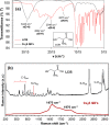
Fig. 2. (a) FTIR spectra comparison of LCIS and Cu2S QD's, (b) Raman spectra of LCIS and Cu2S QD's, a visual reference of the structure of LCIS used isomer is shown as insets.
Fig. 3
Fig. 3. XRD pattern of the Cu2S QD's and comparison with diffraction planes of reported Cu2S cubic phase.
Fig. 4
Fig. 4. UV-vis spectra of Cu2S QD's, a wide absorption behavior is seen ranging from UV to near-infrared.
Fig. 5
Fig. 5. Emission (red line) and emission (black line) fluorescence spectra of LCIS functionalized Cu2S QD's.
Fig. 6
Fig. 6. STEM images of the Cu2S QD's at (a) 500 00×, (b) 1 000 00×, (c) photograph of Cu2S QD's suspension two weeks after it’s synthesis (d) example of the temperature effect in size distribution of Cu2S nanocrystals reported by Du et al.
Fig. 7
Fig. 7. TEM image of Cu2S QDs (a), EDS elemental mapping of the image (a) for Cu (b), and for S (c).
Fig. 8
Fig. 8. EDS analysis of functionalized Cu2S QDs.
Fig. 9
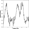
Fig. 9. Profile of diameters of the QD's obtained with TEM.
Fig. 10
Fig. 10. Influence of Cu : S molar ratio in fluorescence emission intensity, crystallite size XRD results for LCIS : Cu and Cu : S molar ratio variations included as table insets.
Fig. 11
Fig. 11. As(iii) detection test following the change in fluorescent emission: before As(iii) addition (black line) and after As(iii) addition (red line).

References

    1. Kang M. Hua Z. Zhenglu W. Haorui C. Kuankuan Z. Xiqing L. Zhugen Y. Biosens. Bioelectron. 2020;148:111785. doi: 10.1016/j.bios.2019.111785. - DOI - PubMed
    1. Ali E. Nafiseh K. Behzad R. Biosens. Bioelectron. 2016;77:499. doi: 10.1016/j.bios.2015.10.011. - DOI - PubMed
    1. Olga L. Rogelio C. Nadia P. Yolanda A. Melissa B. Diana O. Rev. Int. Contam. Ambient. 2017;33:281. doi: 10.20937/RICA.2017.33.02.09. - DOI
    1. Michael F. Siegfried K. Claudia B. Nina H. Stefano B. Micheline K. Mutat. Res., Rev. Mutat. Res. 2020;786:108342. doi: 10.1016/j.mrrev.2020.108342. - DOI - PubMed
    1. Madhusnata D. Tulika C. Drug Chem. Toxicol. 2009;32:169. doi: 10.1080/01480540802594509. - DOI - PubMed