

Histopathological autopsy findings in lungs of pregnant and postpartum women who died of COVID-19 infections
- PMID: 37363653
- PMCID: PMC10078060
- DOI: 10.1007/s00194-023-00618-z
Histopathological autopsy findings in lungs of pregnant and postpartum women who died of COVID-19 infections
Abstract
Background: The coronavirus disease 2019 outbreak (COVID-19) caused by the SARS-CoV coronavirus, has been declared as a pandemic by the World Health Organization on 11 March 2020, as a result of which about 315,000,000 people all over the world have been infected and more than 5,000,000 died.
Objective: Many scientific articles have been published concerning histopathological changes in different organs, but data concerning the lung changes of pregnant and postpartum period women who died of COVID-19 infections are still scarce. The aim of our study was to review and summarize autopsy findings and histopathological changes in lungs of pregnant and postpartum period women who died of COVID-19 infections in Armenia during 2020-2021.
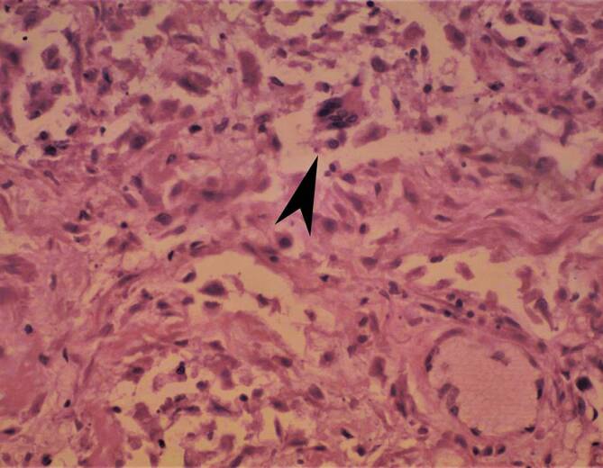
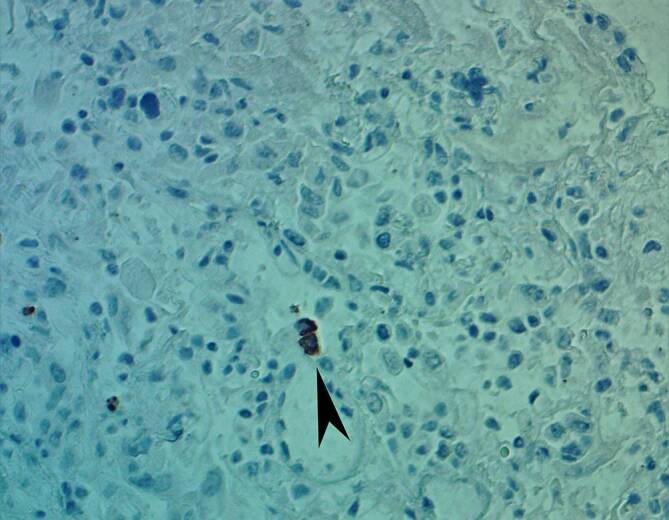
Material and methods: Lung tissue specimens of 14 pregnant and postpartum period women who died of COVID-19 infections and its complications were examined. Hematoxylin-eosin and van Gieson staining methods as well as immunohistochemical examinations were used.
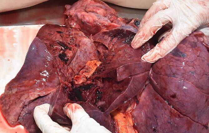
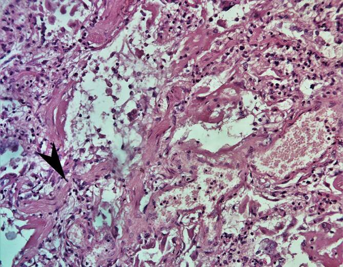
Results: The average age of the dead women was 33.9 years. From 14 cases in 9 there were comorbidities. All cases of death were in the 2nd and 3rd trimester of pregnancy or early and late postpartum period. Forensic medical diagnosis included COVID-19 infection with bilateral polysegmented pneumonia and acute respiratory failure. Histopathological examination revealed diffuse alveolar damage of lungs (DAD) in predominantly proliferation/organizing stage.
Conclusion: Histopathological examination of lungs showed proliferative stage of DAD with signs of fibrosing pneumonia. Early diagnosis and hospitalization of pregnant women may prevent late complications of COVID-19 infection and fibrosing pneumonia development, as well as future risks of fatal outcomes in pregnant and early postpartum period.
Grundlagen: Der Ausbruch der Coronavirus-Krankheit 2019 (COVID-19), verursacht durch das Coronavirus SARS-CoV‑2, wurde am 11. März 2020 von der Weltgesundheitsorganisation zu einer Pandemie erklärt. Als Folge infizierten sich etwa 315.000.000 Menschen auf der ganzen Welt, von denen mehr als 5.000.000 starben.
Ziel: Es wurden viele wissenschaftliche Artikel über histopathologische Veränderungen in verschiedenen Organen veröffentlicht, aber Daten über die Lungenveränderungen von schwangeren und postpartalen Frauen, die an einer COVID-19-Infektion verstarben, sind noch rar. Das Ziel unserer Studie war es, die Autopsiebefunde und histopathologischen Veränderungen in der Lunge von Frauen in der Schwangerschaft und nach der Geburt, die im Zeitraum 2020–2021 in Armenien an einer COVID-19-Infektion starben, zu überprüfen und zusammenzufassen.
Materialien und methoden: Es wurden Lungengewebeproben von 14 schwangeren und postpartalen Frauen entnommen, die an einer COVID-19-Infektion starben, und deren Komplikationen untersucht. Es wurden Hämatoxylin-Eosin und die Färbungsmethode nach van Gieson sowie eine immunhistochemische Untersuchung verwendet.
Ergebnisse: Das Durchschnittsalter der toten Frauen betrug 33,9 Jahre. In 9 von 14 Fällen lagen Komorbiditäten vor. Alle Todesfälle traten im 2. und 3. Trimenon der Schwangerschaft oder im frühen und späten Wochenbett auf. Die gerichtsmedizinische Diagnose umfasste eine COVID-19-Infektion mit bilateralen polysegmentierten Pneumonien und akutem Lungenversagen. Die histopathologische Untersuchung ergab eine diffuse alveoläre Schädigung der Lunge (DAD) im überwiegenden Proliferations‑/Organisierungsstadium.
Schlussfolgerung: Die histopathologische Untersuchung der Lungen zeigte ein proliferatives Stadium der DAD mit Anzeichen einer fibrosierenden Pneumonie. Eine frühzeitige Diagnose und Krankenhauseinweisung schwangerer Frauen könnte Spätkomplikationen einer COVID-19-Infektion und die Entwicklung einer fibrosierenden Lungenentzündung sowie zukünftige Risiken tödlicher Ausgänge bei Schwangeren und in der frühen Zeit nach der Geburt verhindern.
Keywords: Autopsy; COVID-19; Lungs; Morphology; Pregnant women.
© The Author(s), under exclusive licence to Springer Medizin Verlag GmbH, ein Teil von Springer Nature 2023.
Conflict of interest statement
Conflict of interestM.S. Bisharyan, K.A. Arsenyan, P.S. Khachatryan, M.Z. Muradyan and A.A. Tonoyan declare that they have no competing interests.
Figures


References
LinkOut - more resources
Full Text Sources
Miscellaneous