A non-coding insertional mutation of Grhl2 causes gene over-expression and multiple structural anomalies including cleft palate, spina bifida and encephalocele
- PMID: 37364051
- PMCID: PMC10460492
- DOI: 10.1093/hmg/ddad094
A non-coding insertional mutation of Grhl2 causes gene over-expression and multiple structural anomalies including cleft palate, spina bifida and encephalocele
Abstract
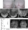
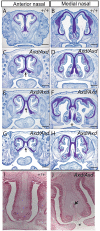
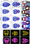
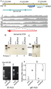
Orofacial clefts, including cleft lip and palate (CL/P) and neural tube defects (NTDs) are among the most common congenital anomalies, but knowledge of the genetic basis of these conditions remains incomplete. The extent to which genetic risk factors are shared between CL/P, NTDs and related anomalies is also unclear. While identification of causative genes has largely focused on coding and loss of function mutations, it is hypothesized that regulatory mutations account for a portion of the unidentified heritability. We found that excess expression of Grainyhead-like 2 (Grhl2) causes not only spinal NTDs in Axial defects (Axd) mice but also multiple additional defects affecting the cranial region. These include orofacial clefts comprising midline cleft lip and palate and abnormalities of the craniofacial bones and frontal and/or basal encephalocele, in which brain tissue herniates through the cranium or into the nasal cavity. To investigate the causative mutation in the Grhl2Axd strain, whole genome sequencing identified an approximately 4 kb LTR retrotransposon insertion that disrupts the non-coding regulatory region, lying approximately 300 base pairs upstream of the 5' UTR. This insertion also lies within a predicted long non-coding RNA, oriented on the reverse strand, which like Grhl2 is over-expressed in Axd (Grhl2Axd) homozygous mutant embryos. Initial analysis of the GRHL2 upstream region in individuals with NTDs or cleft palate revealed rare or novel variants in a small number of cases. We hypothesize that mutations affecting the regulation of GRHL2 may contribute to craniofacial anomalies and NTDs in humans.
© The Author(s) 2023. Published by Oxford University Press.
Figures


Similar articles
-
Over-expression of Grhl2 causes spina bifida in the Axial defects mutant mouse.Hum Mol Genet. 2011 Apr 15;20(8):1536-46. doi: 10.1093/hmg/ddr031. Epub 2011 Jan 24. Hum Mol Genet. 2011. PMID: 21262862 Free PMC article.
-
Shared molecular networks in orofacial and neural tube development.Birth Defects Res. 2017 Jan 30;109(2):169-179. doi: 10.1002/bdra.23598. Birth Defects Res. 2017. PMID: 27933721
-
Overexpression of Grainyhead-like 3 causes spina bifida and interacts genetically with mutant alleles of Grhl2 and Vangl2 in mice.Hum Mol Genet. 2018 Dec 15;27(24):4218-4230. doi: 10.1093/hmg/ddy313. Hum Mol Genet. 2018. PMID: 30189017 Free PMC article.
-
Grainyhead-like Transcription Factors in Craniofacial Development.J Dent Res. 2017 Oct;96(11):1200-1209. doi: 10.1177/0022034517719264. Epub 2017 Jul 11. J Dent Res. 2017. PMID: 28697314 Review.
-
MicroRNAs and Gene Regulatory Networks Related to Cleft Lip and Palate.Int J Mol Sci. 2023 Feb 10;24(4):3552. doi: 10.3390/ijms24043552. Int J Mol Sci. 2023. PMID: 36834963 Free PMC article. Review.
Cited by
-
Elevated GRHL2 Imparts Plasticity in ER-Positive Breast Cancer Cells.Cancers (Basel). 2024 Aug 21;16(16):2906. doi: 10.3390/cancers16162906. Cancers (Basel). 2024. PMID: 39199676 Free PMC article.
-
Meta-analysis reveals transcription factors and DNA binding domain variants associated with congenital heart defect and orofacial cleft.medRxiv [Preprint]. 2025 Feb 2:2025.01.30.25321274. doi: 10.1101/2025.01.30.25321274. medRxiv. 2025. PMID: 39974057 Free PMC article. Preprint.
References
-
- Stanier, P. and Moore, G.E. (2004) Genetics of cleft lip and palate: syndromic genes contribute to the incidence of non-syndromic clefts. Hum. Mol. Genet., 13, 73R–781R. - PubMed
Publication types
MeSH terms
Substances
Grants and funding
LinkOut - more resources
Full Text Sources
Medical
Molecular Biology Databases
Miscellaneous

