. 2023 Jul 4;120(27):e2302534120.
doi: 10.1073/pnas.2302534120. Epub 2023 Jun 26.

Antisense oligonucleotide rescue of CGG expansion-dependent FMR1 mis-splicing in fragile X syndrome restores FMRP

Affiliations

Antisense oligonucleotide rescue of CGG expansion-dependent FMR1 mis-splicing in fragile X syndrome restores FMRP

Sneha Shah et al. Proc Natl Acad Sci U S A. .

Abstract

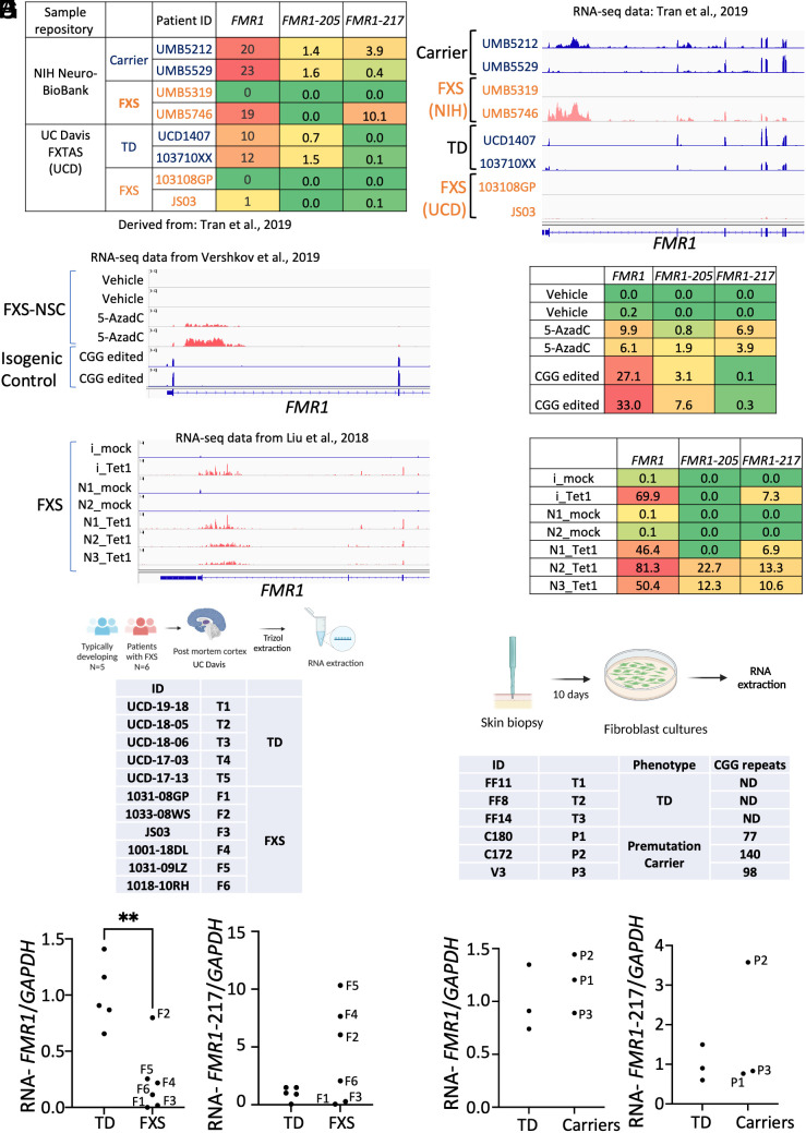
Aberrant alternative splicing of mRNAs results in dysregulated gene expression in multiple neurological disorders. Here, we show that hundreds of mRNAs are incorrectly expressed and spliced in white blood cells and brain tissues of individuals with fragile X syndrome (FXS). Surprisingly, the FMR1 (Fragile X Messenger Ribonucleoprotein 1) gene is transcribed in >70% of the FXS tissues. In all FMR1-expressing FXS tissues, FMR1 RNA itself is mis-spliced in a CGG expansion-dependent manner to generate the little-known FMR1-217 RNA isoform, which is comprised of FMR1 exon 1 and a pseudo-exon in intron 1. FMR1-217 is also expressed in FXS premutation carrier-derived skin fibroblasts and brain tissues. We show that in cells aberrantly expressing mis-spliced FMR1, antisense oligonucleotide (ASO) treatment reduces FMR1-217, rescues full-length FMR1 RNA, and restores FMRP (Fragile X Messenger RibonucleoProtein) to normal levels. Notably, FMR1 gene reactivation in transcriptionally silent FXS cells using 5-aza-2'-deoxycytidine (5-AzadC), which prevents DNA methylation, increases FMR1-217 RNA levels but not FMRP. ASO treatment of cells prior to 5-AzadC application rescues full-length FMR1 expression and restores FMRP. These findings indicate that misregulated RNA-processing events in blood could serve as potent biomarkers for FXS and that in those individuals expressing FMR1-217, ASO treatment may offer a therapeutic approach to mitigate the disorder.

Keywords: FMR1; FMRP; RNA splicing; antisense oligonucleotides.


Conflict of interest statement

S.S., E.B.-K., and J.D.R., have filed a patent on RNA-based biomarkers for Fragile X associated disorders. S.S., J.K.W., and J.D.R. have filed a patent on ASO based therapeutics for Fragile X associated disorders.

Figures

Fig. 1.
Fig. 1.
Gene expression changes in leukocytes derived from FXS individuals and identification of a truncated FMR1 RNA transcript. (A) Schematic diagram of leukocyte isolation from fresh blood samples from FXS male (N = 29) and age-matched TD male (N = 13) individuals and subsequent RNA-seq. The data were analyzed for changes in differential gene expression (DGE), and differential alternative splicing (DAS). (B) Summary table for changes in alternative splicing events in FXS vs. TD leukocytes detected by rMATS (15) at an FDR < 5% and a difference in the exon inclusion levels (PSI, Percent spliced-in) between the genotypes (deltaPSI) of ≥5%. Schematic for the splicing event categories is shown at the left of the table (see also Dataset S2). (C) Violin plots of alternative splicing in FXS vs. TD leukocytes indicating PSI for each type event. (D) RT-PCR showing exon 3 skipping of the LAIR2 RNA in TD (N = 3) and FXS (N = 9) samples. (E) Normalized gene counts (transcripts per million, TPM) obtained from RNA-seq data analysis for total FMR1 (all isoforms), FMR1-205 (encodes full-length 632 amino acid FMRP), FMR1-217 (a mis-spliced RNA, see panel G), and FXR2, a paralog of FMR1. The color scale from red to green denotes highest to lowest gene counts. (F) IGV viewer tracks of RNA-seq data for FXS and TD individuals for the FMR1 gene. FMR1 RNA is detected in all TD individuals (blue) and FXS individuals 1-21 (coral). The black box marked on the FMR1 gene illustrated at the bottom shows the region of intron 1 with differential reads between TD (1-13) and FXS (1-21) individuals. Expanded view of this region is shown in the bottom. (G) An expanded view of the FMR1 RNA marked with a black box in G. The reads map to an exon that comprises the annotated FMR1-217 isoform. Sequence data for FMR1-217 PCR fragments from FXS RNA sample are shown in SI Appendix, Fig. S1G. H refers to high and L refers to low FMR1.
Fig. 2.
Fig. 2.
Correlation of FXS molecular parameters with IQ. (A) FMR1 gene methylation (in percent as determined by PCR analysis, MPCR), FMRP levels (ng/μg total protein), CGG repeat number, FMR1 (all isoforms, TPM), FMR1-217 (TPM), and full-length FMR1-205 (TPM) in leukocytes are listed as well as IQ (Stanford–Binet) for each FXS individual. N/A, not available (see also Dataset S1). (B) Correlation coefficients for pairwise comparisons for each parameter (less CGG expansion and methylation) listed in panel A. Correlations were based on parameters from 19 FXS individuals because some data were not available from all the 29 samples. (C) 3-dimensional comparison of all parameters (less CGG expansion and methylation) listed in panel A. The color from green to red represents increasing FMR1-205 levels (A). The illustration is based on parameters from 19 FXS individuals because some data were not available from all the 29 samples. (D) FMR1-217 RNA levels (TPM) were assessed between FXS samples with CGG repeat number mosaicism (N = 8) or without (full expansion only) (N = 21). (E) FMR1-217 RNA levels (TPM) were assessed between FXS samples with methylation mosaicism (N = 7) or without (full expansion only) (N = 13) (*P < 0.05, t test).
Fig. 3.
Fig. 3.
FMR1-217 is derived from FMR1, requires the CGG expansion, and is expressed in human postmortem brain tissues (FXS and premutation carriers) and in skin-derived fibroblasts (premutation carrier). (A) Sample information for postmortem FXS frontal cortex and premutation FXS carriers and TD individuals (derived from ref. 17). RNA-seq datasets GSE107867 (NIH samples) and GSE117776 were reanalyzed for DGE and DAS. The TPM for FMR1 RNA in the samples is shown. (B) IGV tracks of RNA-seq data (17) for FXS and TD individuals for the FMR1 gene. (C) IGV tracks of selected regions of FMR1 reanalyzed from the RNA-seq data of Vershkov et al. (18), who deleted the FMR1 CGG expansion by CRISPR/Cas9 gene editing. Biologic duplicate of iPSC-derived neural stem cells (NSCs) from FXS individuals (FXS-NSC) treated with vehicle or 5-AzadC and isogenic CGG-edited samples are shown. FMR1-217 reads (coral) are detected only in the 5-AzadC-treated samples. (D) Total FMR1, full-length FMR1-205, and FMR1-217 reads (TPM) of the samples noted in panel C are listed. (E) IGV tracks of selected regions of FMR1 reanalyzed from the RNA-seq data of Liu et al. (19), who performed targeted FMR1 gene demethylation in FXS iPSCs and iPSC-derived neurons. FXS iPSCs/iPSC-derived neurons incubated a mock guide RNA (blue tracks) (i_mock or N1_mock, N2_mock) or an FMR1 guide RNA (coral tracks) and Cas9 fused to the Tet1 demethylase (i_Tet1 or N1_Tet1, N2_Tet1, N3_Tet1). (F) Total FMR1, full-length FMR1-205, and FMR1-217 reads (TPM) of the samples noted in panel E are listed. (G) Experimental design of RNA extraction from postmortem cortical tissue obtained from six FXS males (F1 to F6) and five TD (T1 to T5) age-matched males. RT-qPCR data for cortical tissue–derived RNA samples representing abundance for FMR1 and FMR1-217 isoforms relative to GAPDH RNA. Each sample was analyzed in duplicate. Primers used for amplification are represented in SI Appendix, Fig. S1F (**P < 0.01, t test). (H) Schematic diagram of fibroblast generated from skin biopsies obtained from three male premutation carriers (P1 to P3) and three male TD individuals (T1 to T3). The table shows patient deidentified designation, genotypes, and CGG repeat numbers in the 5′UTR in the FMR1 gene. ND, not determined. qPCR data for fibroblast-derived RNA samples representing abundance for FMR1 and FMR1-217 isoforms relative to GAPDH RNA are shown. Each sample was analyzed in duplicate. Primers used for amplification are represented in SI Appendix, Fig. S1F and Dataset S1.
Fig. 4.
Fig. 4.
ASOs targeting FMR1-217 restore FMRP levels in FXS2 LCLs with incomplete methylation. (A) Sample information for lymphoblast cell lines (LCLs) (Coriell Institute, NJ) from two FXS and two TD members of a family is shown. FMRP and GAPDH (loading control) levels were determined by western blots. Ratios of FMRP/GAPDH normalized to FXS1 are shown below the blot. FMRP quantification by Luminex Microplex immunochemistry assay is shown in ng FMRP/μg total protein. (B) The proportion of full-length FMR1 to FMR1-217 was quantified using RT-qPCR in the TD and FXS2 LCLs relative to GAPDH RNA levels. Primers used for q-PCR are shown in the gene illustration. The total FMR1 RNA was unchanged, but the proportion of FMR1-217 was significantly higher in FXS2 LCL compared to TD LCL. (C) ASOs complementary to FMR1-217 RNA are illustrated (intron specific: 704 to 706, intron–exon junction specific: 707 to 710 and exon specific: 711 to 714). (D) Schematic diagram of the ASO treatment (80 nM for 72 h) of the FXS2 LCLs to determine FMR1 isoform and FMRP levels after demethylation. DMSO-treated cells were used as a vehicle control (****P < 0.0001, **P < 0.01, t test). (E) FMRP levels were determined for FXS2 LCLs treated with DMSO (vehicle) and ASOs as described in Fig. 5A. TD LCLs were also probed for FMRP on the same western blots. Ratios of FMRP/GAPDH normalized to FXS1 are shown below the blot. (F) Model depicting that ASO-mediated decrease of mis-spliced FMR1-217 in FXS2 LCLs can restore FMR1 full-length RNA and consequently FMRP levels.
Fig. 5.
Fig. 5.
ASOs targeting FMR1-217 in combination with 5-AzadC restores FMRP levels in FXS cells with complete FMR1 gene methylation. (A) Schematic diagram of the fully methylated FXS cells treated with ASOs 713 and 714 (80 nM each) followed by 5-AzadC (1 μM) added on consecutive days 2 to 9 after which RNA and protein were extracted. (B) FMR1-217 and FMR1 isoforms were assessed using qPCR primers as shown in SI Appendix, Fig. S1F and were analyzed using one-way ANOVA with multiple comparisons test (****P < 0.0001, ***P < 0.001, **P < 0.01, *P < 0.05). Data information: bar graphs indicate mean, and error bars indicate ± SEM. (C) Western blot of FMRP and GAPDH from FXS1 LCLs treated with DMSO, 5-AzadC, or 5-AzadC plus ASOs. Histogram depicting quantification of western blot for FXS1 cells treated with DMSO, 5-AzadC, and ASO or 5-AzadC alone (N = 3) in arbitrary units. Significance was determined using one-way ANOVA with multiple comparisons test (****P < 0.0001). Data information: bar graphs indicate mean, and error bars indicate ± SEM. (D) Lung fibroblasts derived from an FXS individual (GM07072, Coriell Institute) were cultured with 5-AzadC for 8 d and then treated with ASOs 713/714 (100 nM each) for 72 h prior to RNA and protein extraction. RT-qPCR analysis of FMR1-217 FMR1, and GAPDH RNAs in lung fibroblasts treated with DMSO, ASOs 713/714, 5-AzadC, or the ASOs 713/714 plus 5-AzadC. The amounts of FMR1-217 and FMR1 were made relative to GAPDH (*P < 0.05, **P < 0.01, ***P < 0.001, one-way ANOVA with multiple comparisons test). (E) Western blots of FMRP and GAPDH from the lung fibroblasts treated as in panel B. Quantification of FMRP relative to GAPDH is noted below (N = 2) in arbitrary units. Significance was determined using one-way ANOVA with multiple comparisons test (****P < 0.0001, ***P < 0.001, **P < 0.01, one-way ANOVA with multiple comparisons test). Data information: bar graphs indicate mean, and error bars indicate ± SEM. (F) Model depicting active FMR1 transcription in FXS cells (or after treatment with demethylating agents to activate FMR1 transcription) results in the production of mis-spliced FMR1-217. Downregulation of FMR1-217 with an ASO results in rescue of correctly spliced FMR1 transcripts and restoration of FMRP.

References

    1. Darnell J. C., et al. , FMRP stalls ribosomal translocation on mRNAs linked to synaptic function and autism. Cell 14, 247–261 (2011), https://doi.org/10.1016/j.cell.2011.06.013. - DOI - PMC - PubMed
    1. Maurin T., et al. , HITS-CLIP in various brain areas reveals new targets and new modalities of RNA binding by fragile X mental retardation protein. Nucleic Acids Res. 46, 6344–6355 (2018). - PMC - PubMed
    1. Li M., Identification of FMR1-regulated molecular networks in human neurodevelopment. Genome Res. 30, 361–374 (2020), 10.1101/gr.251405.119. - DOI - PMC - PubMed
    1. Sharma S. D., et al. , Widespread alterations in translation elongation in the brain of Juvenile Fmr1 knockout mice. Cell Rep. 26, 3313–3322.e5 (2019), 10.1016/j.celrep.2019.02.086. - DOI - PMC - PubMed
    1. Udagawa T., et al. , Genetic and acute CPEB1 depletion ameliorate fragile X pathophysiology. Nat. Med. 19, 1473 (2013). - PMC - PubMed

Publication types

MeSH terms