. 2023 Jul 13;10(7):ENEURO.0043-23.2023.
doi: 10.1523/ENEURO.0043-23.2023. Print 2023 Jul.

A Cre Driver Line for Genetic Targeting of Kappa Opioid Receptor Expressing Cells

Affiliations

A Cre Driver Line for Genetic Targeting of Kappa Opioid Receptor Expressing Cells

Franciely Paliarin et al. eNeuro. .

Abstract

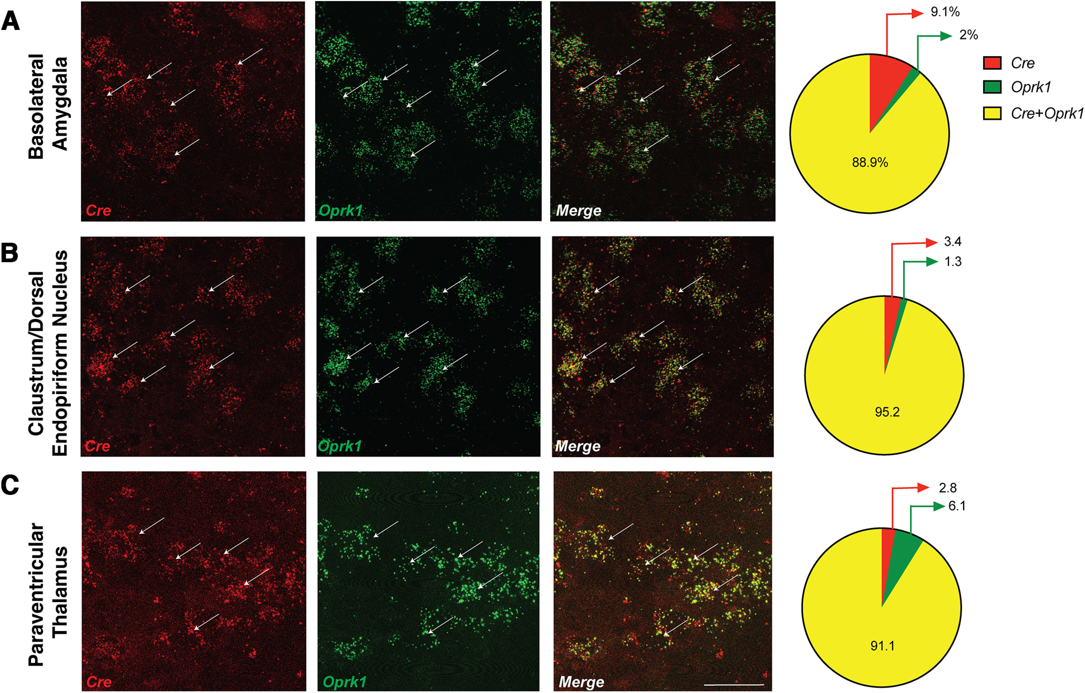
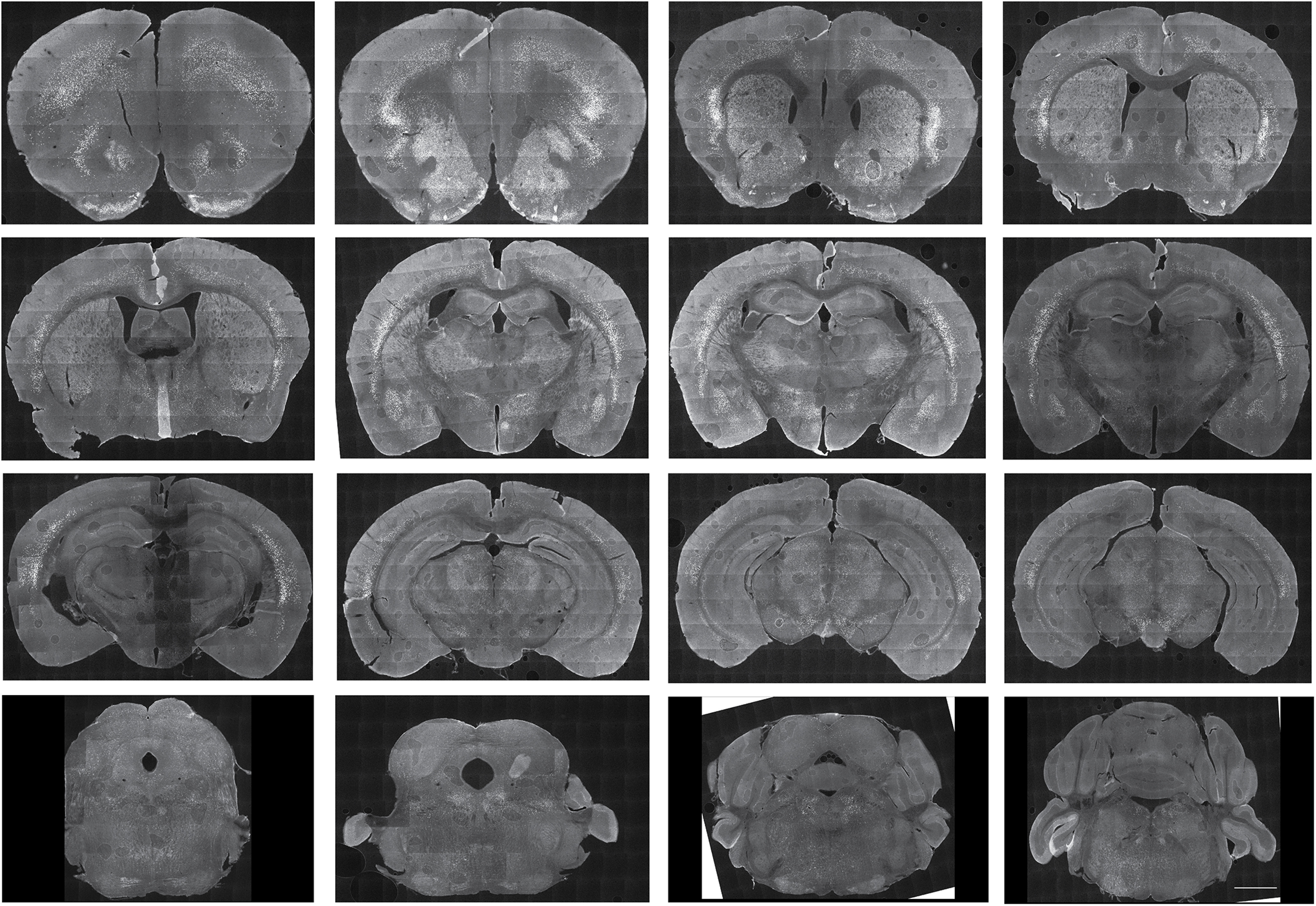
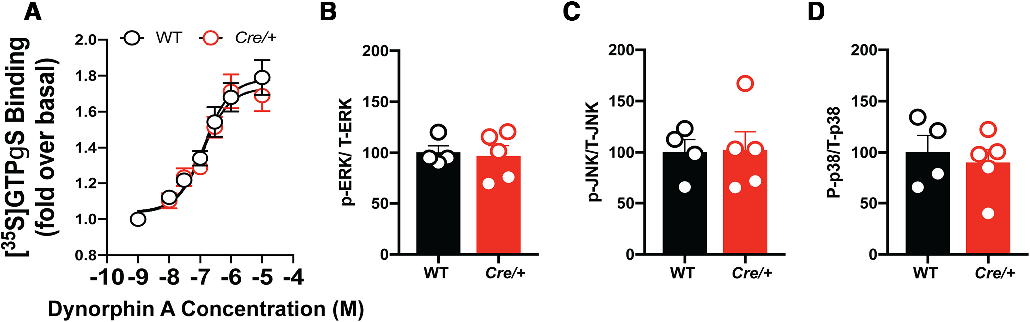
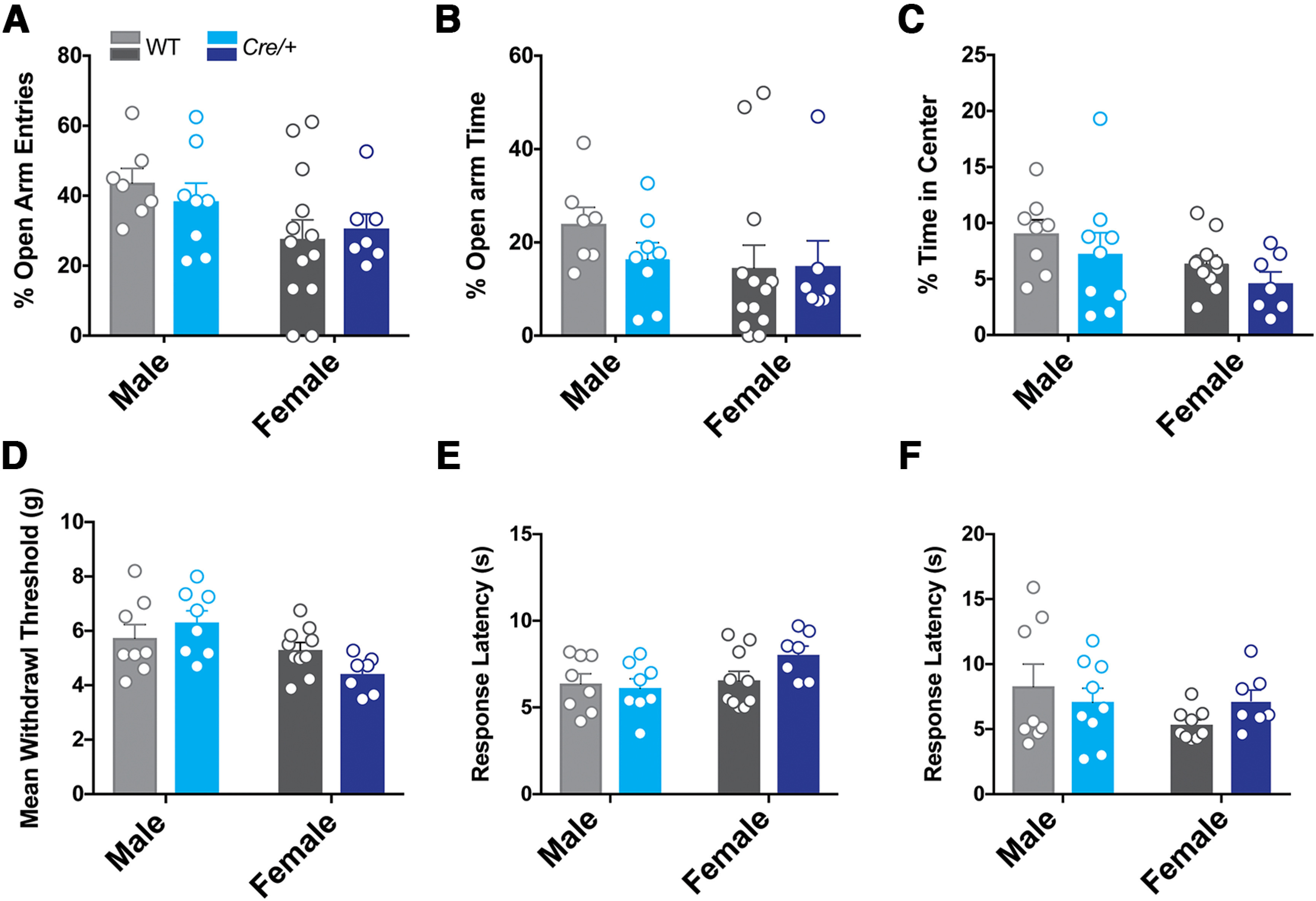
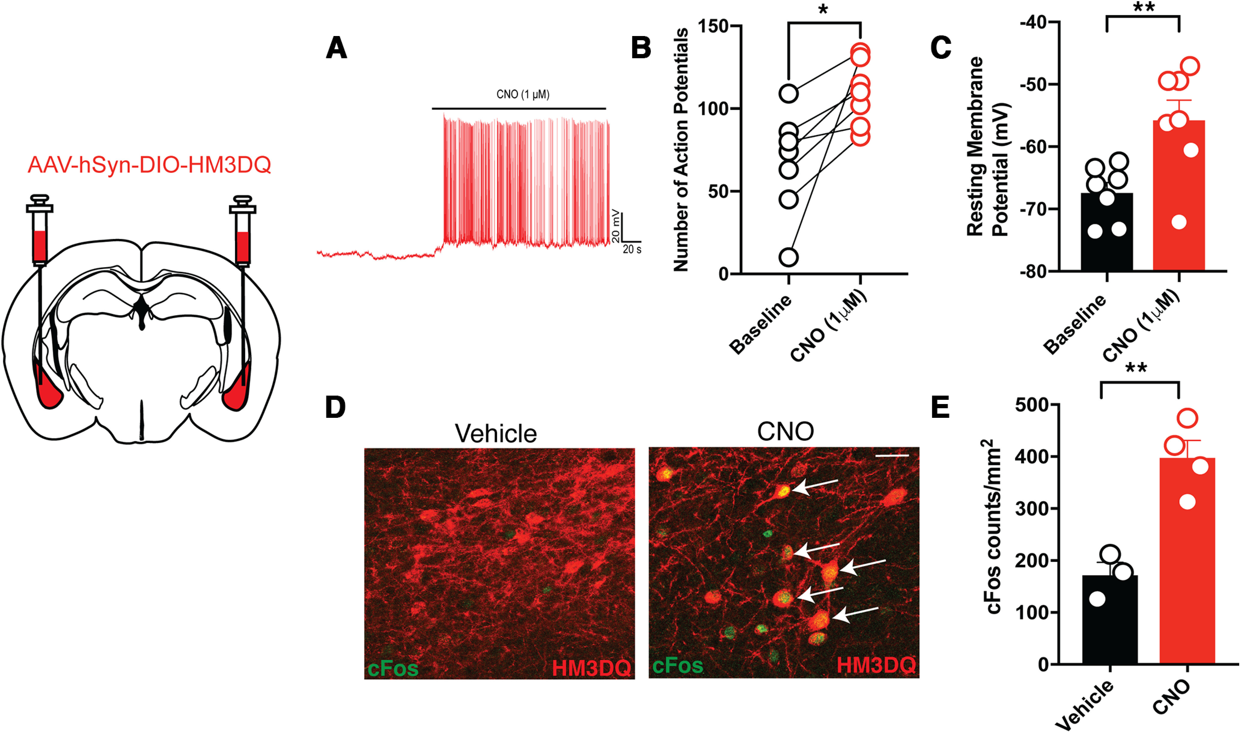
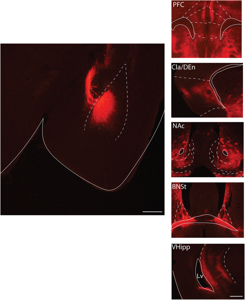
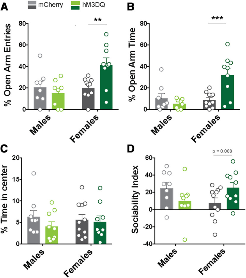
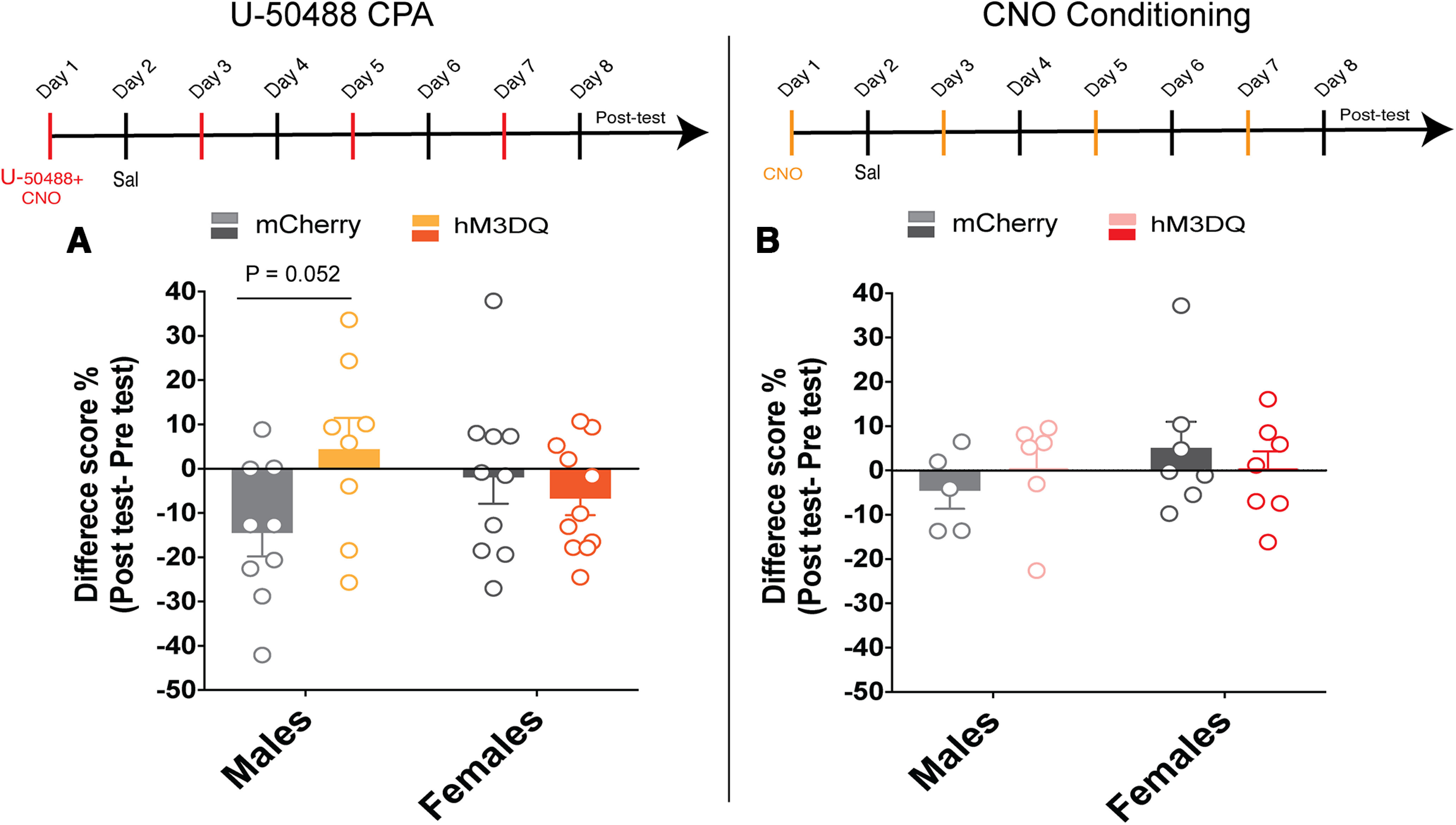
Here we describe the generation and characterization of a Cre knock-in mouse line that harbors a Cre insertion in the 3'UTR of the κ opioid receptor gene (Oprk1) locus and provides genetic access to populations of κ opioid receptor (KOR)-expressing neurons throughout the brain. Using a combination of techniques including RNA in situ hybridization and immunohistochemistry, we report that Cre is expressed with high fidelity in KOR-expressing cells throughout the brain in this mouse line. We also provide evidence that Cre insertion does not alter basal KOR function. Baseline anxiety-like behaviors and nociceptive thresholds are unaltered in Oprk1-Cre mice. Chemogenetic activation of KOR-expressing cells in the basolateral amygdala (BLAKOR cells) resulted in several sex-specific effects on anxiety-like and aversive behaviors. Activation led to decreased anxiety-like behavior on the elevated plus maze and increased sociability in female but not in male Oprk1-Cre mice. Activation of BLAKOR cells also attenuated KOR agonist-induced conditioned place aversion (CPA) in male Oprk1-Cre mice. Overall, these results suggest a potential role for BLAKOR cells in regulating anxiety-like behaviors and KOR-agonist mediated CPA. In summary, these results provide evidence for the utility of the newly generated Oprk1-Cre mice in assessing localization, anatomy, and function of KOR circuits throughout the brain.

Keywords: anxiety; conditioned place aversion; dynorphin; genetic access; knock-in mice; social interaction.


Figures

Figure 1.
Figure 1.
Colocalization of Oprk1 and Cre. AC, In situ hybridization using probes targeting Cre recombinase and mouse Oprk1 reveal 80–90% colocalization between Oprk1 and Cre transcripts across the following three brain regions: the BLA (A), claustrum/dorsal endopiriform nucleus (B), and PVT (C). N = 2 male mice; N = 1 female mouse. Scale bar, 50 µm. See Extended Data Figure 1-1 for generation of Oprk1-Cre transgenic mouse.
Figure 2.
Figure 2.
Brain-wide snapshots of Cre expression in the adult Oprk1-Cre mouse. Tiled images of Cre protein expression at 20× resolution across the brain of an adult Oprk1-Cre mouse are shown. Strongest Cre expression was observed in the claustrum and dorsal endopiriform nucleus. Cre expression was also observed in the PVT, CeA, BLA, PVT, DR, and locus coeruleus (LC). The pattern of Cre expression matched that of Oprk1 expression in the Allen Brain Atlas. Scale bar, 1 mm. Please see Extended Data Figure 2-1 for distribution of BLAKOR cells and Extended Data Figure 2-2 for colocalization between virally delivered Cre-dependent EGFP and Oprk1 in the BLA.
Figure 3.
Figure 3.
KOR function is intact in Oprk1-Cre mice. A, Dyn-stimulated GTPγS binding did not differ significantly between WT and Oprk1-Cre mice. n = 7 males/group. B–D, No differences were observed in phosphorylated (P)-ERK (B), P-JNK (C), and P-p38 (D) in the NAc under basal conditions. N = 4–5/group (WT = 4 females, Cre/+ = 3 females and 2 males). Please see Extended Data Figure 3-1 for Oprk1 mRNA expression in WT and Oprk1-Cre mice; Extended Data Figure 3-2 for [35S]GTPγS binding in the striatum of WT and Oprk1-Cre mice; and Extended Data Figure 3-3 for basal KOR signaling is not altered in the amygdala of Oprk1-Cre mice. Extended Data Table 3-1 lists antibodies used for Western blotting.
Figure 4.
Figure 4.
Baseline anxiety-like and pain behaviors in Oprk1-Cre mice. AC, Anxiety-like behaviors were tested using the EPM and open field anxiety tests in WT and Oprk1-Cre mice. There were no significant genotypic differences in open arm entries (A) or time (B) in the EPM (WT male = 7, WT female = 12; Cre/+ male = 8, Cre/+ female = 7). C, There were also no genotypic differences in the percentage of time in the center of the open field (WT male = 8, WT female = 11; Cre/+ male = 9, Cre/+ female = 7). D, There were no genotype differences seen in the mean withdrawal threshold in the eVF mechanical nociception test (WT male = 8, WT female = 10; Cre/+ male = 8, Cre/+ female = 7) and response latencies (D) on the hot plate tests (E; WT male = 8, WT female = 10; Cre/+ male = 8, Cre/+ female = 7) and cold plate tests (F) and (WT male = 8, WT female = 9; Cre/+ male = 9, Cre/+ female = 7). See Extended Data Figure 4-1 for closed arm entries were not altered on the EPM in Oprk1-Cre mice.
Figure 5.
Figure 5.
Validation of DREADD-induced activation of BLAKOR neurons. AC, CNO application increased both spontaneous and electrically evoked excitability in BLAKOR cells expressing hM3DQ. *p = 0.0217, **p = 0.0091, paired t test, n = 7 cells/group. D, Visual representation of c-Fos activation in the BLA of hM3DQ-injected Oprk1-Cre mice treated with vehicle or CNO before killing. E, Quantification of c-Fos shows a significant increase in the activation in CNO-injected mice compared with vehicle-injected mice. **p = 0.0041, unpaired t test, N =3–4 mice/group.
Figure 6.
Figure 6.
Projections of BLAKOR neurons. Image of mCherry injection into the BLA (left). BLAKOR neurons project to the prefrontal cortex (PFC), the claustrum/dorsal endopiriform nucleus, the NAc, the bed nucleus of the stria terminalis (BNST), and the ventral hippocampus (VHipp; right).
Figure 7.
Figure 7.
Chemogenetic activation of BLAKOR cells and anxiety-like behavior. A, There was no significant difference between mCherry and hM3DQ-injected male mice in open arm entries or open arm time in males (N = 8/group). B, There was a significant increase in the percentage of open arm entries (**p = 0.0016) and open arm time (***p = 0.0010, Sidak post-test) in hM3DQ-injected female mice compared with mCherry controls (N =9–10 mice/group). Please see Extended Data Figure 7-1 for closed arm entries in mCherry-injected and hM3DQ-injected mice. C, There were no differences in the time spent in the center of an open field in male or female hM3DQ-injected mice compared with mCherry controls (mCherry male = 9, mCherry female = 11; hM3DQ male = 9, hM3DQ female = 9). D, There was no difference in sociability index in male mCherry-injected and hM3DQ-injected mice. Female hM3Dq mice showed increased sociability compared with mCherry controls. This result did not reach statistical significance (p = 0.088, Sidak post-test; mCherry male = 9, mCherry female = 11; hM3DQ male = 9, hM3DQ female = 10).
Figure 8.
Figure 8.
Chemogenetic activation of BLAKOR cells attenuates U-50488 induced CPA. A, CNO administration attenuated U-50488-induced CPA in hM3DQ-injected males compared with mCherry controls. However, this effect did not reach statistical significance (p = 0.0524, Sidak post-test; N = 8–9/group) in males and in females (N = 9–11/group). B, Neither male nor female hM3DQ-injected Oprk1-Cre mice form a preference for or avoid the CNO-paired chamber. N = 5–6/group for males and N = 5–7/group for females. See Extended Data Figure 8-1 for U-50488-induced CPA in C57BL/6J mice.

Similar articles

Cited by

References

    1. Abraham AD, Casello SM, Schattauer SS, Wong BA, Mizuno GO, Mahe K, Tian L, Land BB, Chavkin C (2021) Release of endogenous dynorphin opioids in the prefrontal cortex disrupts cognition. Neuropsychopharmacology 46:2330–2339. 10.1038/s41386-021-01168-2 - DOI - PMC - PubMed
    1. Al-Hasani R, Bruchas MR (2011) Molecular mechanisms of opioid receptor-dependent signaling and behavior. Anesthesiology 115:1363–1381. 10.1097/ALN.0b013e318238bba6 - DOI - PMC - PubMed
    1. Bäckman CM, Malik N, Zhang Y, Shan L, Grinberg A, Hoffer BJ, Westphal H, Tomac AC (2006) Characterization of a mouse strain expressing Cre recombinase from the 3′ untranslated region of the dopamine transporter locus. Genesis 44:383–390. 10.1002/dvg.20228 - DOI - PubMed
    1. Becker JB, Chartoff E (2019) Sex differences in neural mechanisms mediating reward and addiction. Neuropsychopharmacology 44:166–183. 10.1038/s41386-018-0125-6 - DOI - PMC - PubMed
    1. Bohn LM, Zhou L, Ho JH (2015) Approaches to assess functional selectivity in GPCRs: evaluating G protein signaling in an endogenous environment. Methods Mol Biol 1335:177–189. - PMC - PubMed

LinkOut - more resources