. 2023 Jun 26;13(1):10311.
doi: 10.1038/s41598-023-37469-3.

Sperm-fluid-cell interplays in the bovine oviduct: glycosaminoglycans modulate sperm binding to the isthmic reservoir

Affiliations

Sperm-fluid-cell interplays in the bovine oviduct: glycosaminoglycans modulate sperm binding to the isthmic reservoir

Coline Mahé et al. Sci Rep. .

Abstract

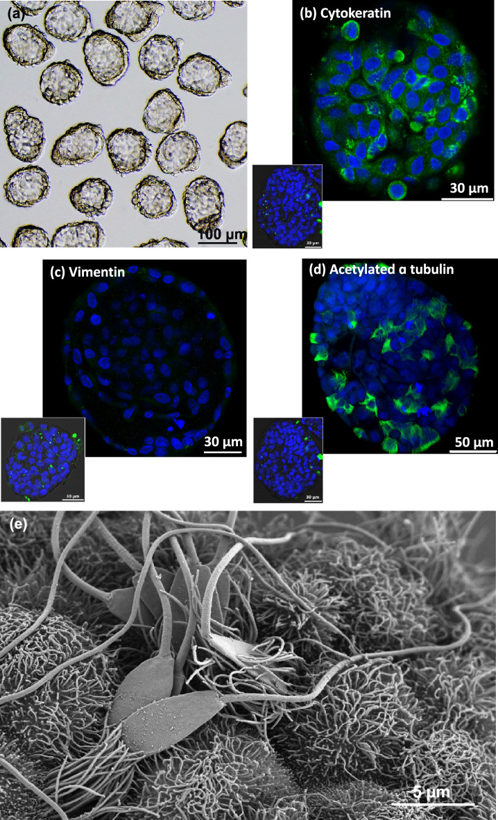
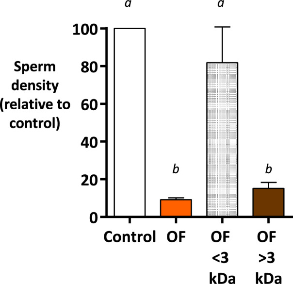
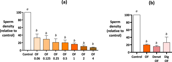
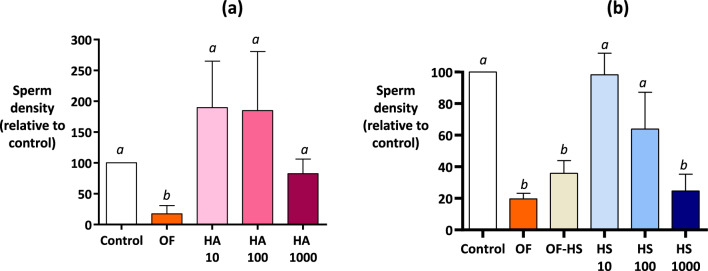
When entering the oviduct for fertilisation, spermatozoa come into contact with the oviduct fluid (OF) and can bind to luminal epithelial cells in the isthmus to form a sperm reservoir. The objective of this study was to examine how the OF modulates sperm adhesion to the oviduct reservoir using an in vitro model of oviduct epithelial spheroids (OES). Bovine oviducts from a local slaughterhouse were used to collect OF and isthmic fragments for the in vitro incubation of OES. Compared to a non-capacitating control medium, the pre-ovulatory OF significantly decreased by 80-90% the density of spermatozoa bound to OES without affecting sperm motility, membrane integrity, or sperm-cilia interactions. This effect on sperm binding was reproduced with (1) OF from different cycle stages and anatomical regions of the oviduct; (2) OF fractions of more than 3 kDa; (3) modified OF in which proteins were denatured or digested and (4) heparan sulphate but not hyaluronic acid, two glycosaminoglycans present in the OF. In conclusion, the OF significantly decreased the number of spermatozoa that bind to oviduct epithelial cells without affecting sperm motility and this effect was due to macromolecules, including heparan sulphate.


Conflict of interest statement

The authors declare no competing interests.

Figures

Figure 1
Figure 1
Characterisation of bovine isthmic epithelial spheroids as an in vitro model to study sperm-oviduct interactions. Spheroids that were uniform in shape and size were selected for sperm co-incubation (a). The spheroid epithelial cells were stained positively for anti-cytokeratin (b, green signal) and negatively for anti-vimentin (c) antibodies (nuclei are stained in blue). The sperm-spheroid complex that was immunostained for acetylated alpha-tubulin (in green) and stained with Hoechst (in blue) showed sperm heads bound to cilia (d). The sperm-spheroid complex observed by scanning electron microscopy showed sperm heads with an intact acrosome firmly attached to the cilia (e). Inserts in (bd) show the negative controls incubated with the immunoglobulin isotype of the primary antibody.
Figure 2
Figure 2
Kinetics of sperm binding to oviduct epithelial spheroids in the Tyrode Lactate Pyruvate (TLP) control medium and in pre-ovulatory oviduct fluid (Experiment 1). (a) The spermatozoa and spheroids were incubated in the control TLP medium (white bars) or pre-ovulatory oviduct fluid (OF) at a final protein concentration of 1 mg/mL (orange bars) for 5, 15, 30. and 60 min. The data are provided as the mean ± SEM of bound sperm per mm2 (n = 3 replicates). The different letters indicate differences between groups. Confocal microscopy pictures of sperm-spheroid complexes after incubation for 60 min in the TLP medium (left) or pre-ovulatory OF at a 1 mg/mL concentration of proteins (right). The sperm nuclei appear in blue.
Figure 3
Figure 3
Effect of the stage of the cycle (a) and anatomical regions (b) from which the oviduct fluid originated on sperm binding to oviduct epithelial spheroids (Experiment 2). The spermatozoa and spheroids were incubated for 60 min in the TLP medium (control) or in the OF collected from whole oviducts at the pre-ovulatory (Pre-ov) or luteal (Luteal) phases of the cycle (a), or from the isthmus (Isth) or ampulla (Amp) at pre-ovulatory (Pre-ov) or post-ovulatory (Post-ov) phases of the cycle. All of the OF samples used had a final protein concentration of 1 mg/mL. The data are provided as the mean ± SEM of bound sperm per mm2 relative to the control (n = 3 replicates).
Figure 4
Figure 4
Effect of ultra-filtrated fractions of pre-ovulatory oviduct fluid on sperm binding to oviduct epithelial spheroids (Experiment 3). The spermatozoa and spheroids were incubated for 60 min with pre-ovulatory OF after ultrafiltration with a cut-off of 3 kDa. Native OF at a final protein concentration of 1 mg/mL was included (OF). The TLP medium was used as the control. The data are presented as the mean ± SEM of bound sperm per mm2 relative to the control (n = 4 replicates).
Figure 5
Figure 5
Effect of proteins in the oviduct fluid on sperm binding to oviduct epithelial spheroids (Experiment 4). The spermatozoa and spheroids were incubated for 60 min in the pre-ovulatory OF at final protein concentrations of 0.06, 0.125, 0.25, 0.5, 1, 2, or 4 mg/mL (a), or in the OF at a final concentration of 1 mg/mL of proteins (OF) and after protein denaturation by heating (Denat OF) or protein digestion by proteinase K treatment (Dig OF). The TLP medium was used as control (a, b). The data are provided as the mean ± SEM of the bound sperm per mm2 relative to control (n = 4 replicates for each experiment).
Figure 6
Figure 6
Effect of hyaluronic acid and heparan sulphate on sperm density on bovine oviduct epithelial spheroids (Experiment 5). The spermatozoa and spheroids were incubated for 60 min with hyaluronic acid (HA) at 10, 100, and 1000 µg/mL (a), or with heparan sulphate (HS) at 10, 100, and 1000 µg/mL and OF in which the HS was digested with heparinases (OF-HS, b). Native OF at a final protein concentration of 1 mg/mL was included (OF) and the TLP medium was used as control in each experiment. The data are presented as the mean ± SEM of bound sperm per mm2 relative to the control (n = 4 replicates for each experiment).

References

    1. Hunter RHF, Nichol R. Transport of spermatozoa in the sheep oviduct: Preovulatory sequestering of cells in the caudal isthmus. J. Exp. Zool. 1983;228:121–128. doi: 10.1002/jez.1402280113. - DOI - PubMed
    1. Lefebvre R, et al. Characterization of the oviductal sperm reservoir in cattle. Biol. Reprod. 1995;53:1066–1074. doi: 10.1095/biolreprod53.5.1066. - DOI - PubMed
    1. Boilard M, Bailey J, Collin S, Dufour M, Sirard M-A. Effect of bovine oviduct epithelial cell apical plasma membranes on sperm function assessed by a novel flow cytometric approach. Biol. Reprod. 2002;67:1125–1132. doi: 10.1095/biolreprod67.4.1125. - DOI - PubMed
    1. Boilard M, et al. Localization of the chaperone proteins GRP78 and HSP60 on the luminal surface of bovine oviduct epithelial cells and their association with spermatozoa. Biol. Reprod. 2004;71:1879–1889. doi: 10.1095/biolreprod.103.026849. - DOI - PubMed
    1. Lamy J, et al. Steroid hormones regulate sperm–oviduct interactions in the bovine. Reproduction. 2017;154:497–508. doi: 10.1530/REP-17-0328. - DOI - PubMed

Publication types