

Influence of posterior staphyloma in myopic maculopathy and visual prognosis
- PMID: 37365301
- PMCID: PMC10764733
- DOI: 10.1038/s41433-023-02648-z
Influence of posterior staphyloma in myopic maculopathy and visual prognosis
Abstract
Background/objectives: Posterior staphyloma is a hallmark of high myopia and its presence associates to greater degrees of myopic maculopathy. Nonetheless, its development, repercussion on visual function and relationship with maculopathy components, is still unclear. The objective was to analyze the impact of posterior staphyloma on the incidence and severity of myopic maculopathy and its repercussion on visual prognosis.
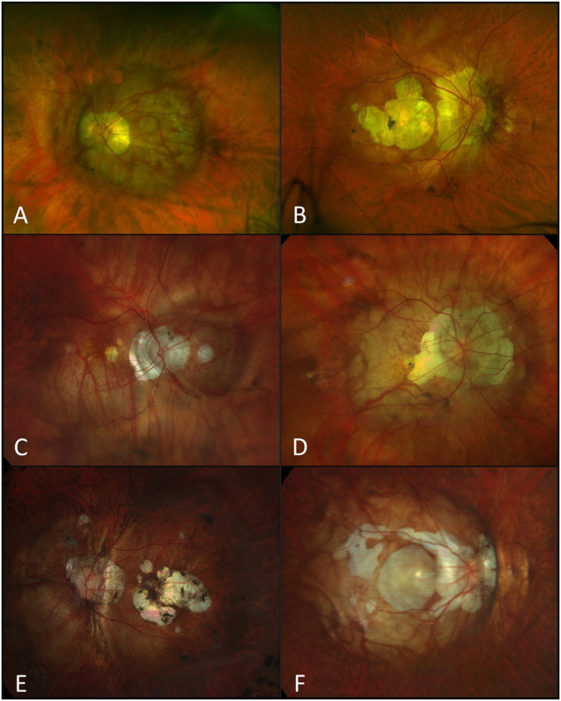
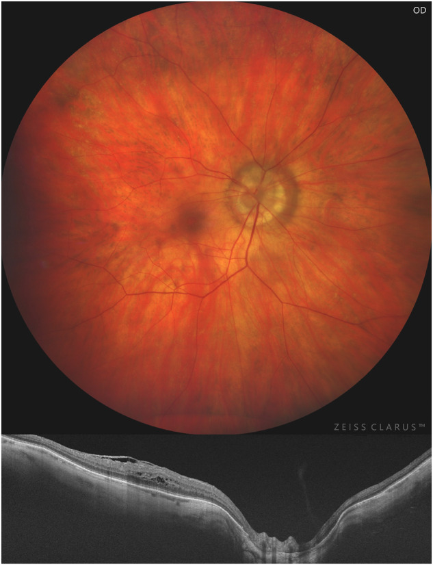
Subjects/methods: Cross-sectional study conducted on 473 consecutive eyes of 259 highly myopic patients examined at Puerta de Hierro-Majadahonda University Hospital (Madrid, Spain). All patients underwent complete ophthalmologic examination including best corrected visual acuity (BCVA), axial length (AL), myopic maculopathy classification according to ATN system (atrophic/traction/neovascularization), determined the presence of posterior staphyloma, pathologic myopia (PM) and severe PM. Multimodal imaging were performed including fundus photography, optical coherence tomography (OCT), OCT-angiography, fundus autofluorescence and/ or fluorescein angiography.
Results: Out of the total, 70.65% were female patients (n = 173/259), mean BCVA was 0.41 ± 0.54 logMAR units and mean AL was 29.3 ± 2.6 mm (26-37.6). Posterior staphyloma was present in 69.4% of eyes. Eyes with posterior staphyloma compared to non-staphyloma were older (p < 0.05), had greater AL (p < 0.01), worse BCVA (p < 0.01) and higher stage in ATN components (p < 0.01). Moreover, compound subgroup showed worse BCVA (p < 0.01) and greater stage in each of the ATN components (p < 0.01). Staphylomas with macular involvement presented worse BCVA (p < 0.01), higher AL (p < 0.01), and greater ATN (p < 0.05). The risk of posterior staphyloma presence in eyes with PM and severe PM eyes was 89.8% and 96.7%, respectively. Posterior staphyloma was the best predictor for BCVA in myopic patients (p < 0.01).
Conclusions: Posterior staphyloma's presence determines high risk of myopic maculopathy and therefore worse visual prognosis, especially those with macular involvement. Posterior staphyloma represented the best predictor for BCVA in highly myopic patients.
© 2023. The Author(s), under exclusive licence to The Royal College of Ophthalmologists.
Conflict of interest statement
The authors declare no competing interests.
Figures
Comment on
-
Myopic maculopathy: Current status and proposal for a new classification and grading system (ATN).Prog Retin Eye Res. 2019 Mar;69:80-115. doi: 10.1016/j.preteyeres.2018.10.005. Epub 2018 Nov 1. Prog Retin Eye Res. 2019. PMID: 30391362 Review.
References
-
- Spaide RF. Staphyloma: Part I. In: Pathologic myopia. New York, NY: Springer; 2014. pp. 167–76.
Publication types
MeSH terms
LinkOut - more resources
Full Text Sources
Medical
