. 2023 Jun 26;21(1):146.
doi: 10.1186/s12915-023-01647-6.

Dopamine modulates the retinal clock through melanopsin-dependent regulation of cholinergic waves during development

Affiliations

Dopamine modulates the retinal clock through melanopsin-dependent regulation of cholinergic waves during development

Chaimaa Kinane et al. BMC Biol. .

Abstract

Background: The mammalian retina contains an autonomous circadian clock that controls various aspects of retinal physiology and function, including dopamine (DA) release by amacrine cells. This neurotransmitter plays a critical role in retina development, visual signalling, and phase resetting of the retinal clock in adulthood. Interestingly, bidirectional regulation between dopaminergic cells and melanopsin-expressing retinal ganglion cells has been demonstrated in the adult and during development. Additionally, the adult melanopsin knockout mouse (Opn4 -/-) exhibits a shortening of the endogenous period of the retinal clock. However, whether DA and / or melanopsin influence the retinal clock mechanism during its maturation is still unknown.

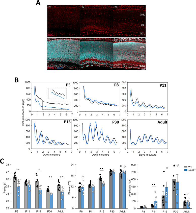
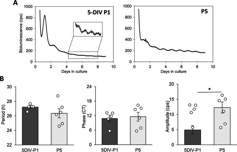
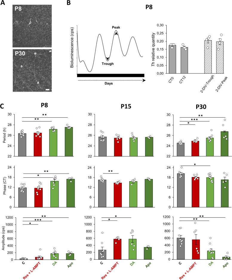
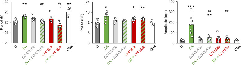
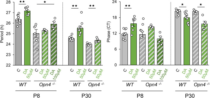
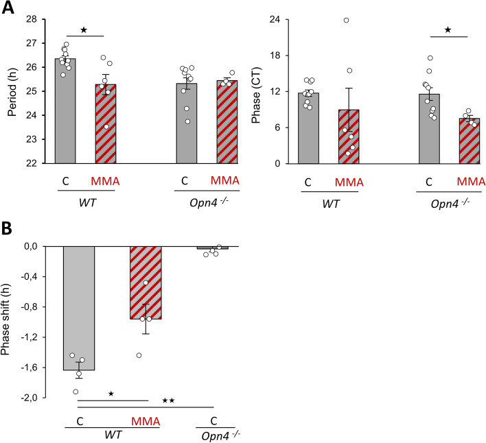
Results: Using wild-type Per2 Luc and melanopsin knockout (Opn4 -/-::Per2 Luc) mice at different postnatal stages, we found that the retina generates self-sustained circadian rhythms from postnatal day 5 in both genotypes and that the ability to express these rhythms emerges in the absence of external time cues. Intriguingly, only in wild-type explants, DA supplementation lengthened the endogenous period of the clock during the first week of postnatal development through both D1- and D2-like dopaminergic receptors. Furthermore, the blockade of spontaneous cholinergic retinal waves, which drive DA release in the early developmental stages, shortened the period and reduced the light-induced phase shift of the retinal clock only in wild-type retinas.

Conclusions: These data suggest that DA modulates the molecular core of the clock through melanopsin-dependent regulation of acetylcholine retinal waves, thus offering an unprecedented role of DA and melanopsin in the endogenous functioning and the light response of the retinal clock during development.

Keywords: Acetylcholine; Circadian rhythms; Dopamine; Light; Melanopsin; Retinal waves.


Conflict of interest statement

The authors declare that they have no competing interests.

Figures

Fig. 1
Fig. 1
Expression of BMAL1 and ontogenesis of PER2::Luc circadian oscillations in wild-type and Opn4 −/− retinal explants. A Immunostaining of BMAL1 (red) in retinal sections of wild-type mice counterstained with DAPI (blue) at P3, P5 and P8. Scale bar = 10 µm: NBL, neuroblastic retina; ONL, outer nuclear layer; INL, inner nuclear layer and GCL, ganglion cell layer. B Representative bioluminescence recording of PER2::Luc retinal explants from Per2 Luc (black line) and Opn4 −/− ::Per2 Luc (blue line) mice at different postnatal stages (P5, P8, P11, P15, P30) and in the adult. Retinal explants were cultured for at least 6 days without medium change. The dotted black rectangle corresponds to an enlargement of bioluminescence traces of both genotypes at P5. C Means of the endogenous period, the circadian phase, and the amplitude in both genotypes during development. The total numbers of retinas used were respectively for wild-type mice: P8, n = 10; P11, n = 10; P15, n = 6; P30, n = 8; adult, n = 10 and for Opn4 −/− ::Per2 Luc; P8, n = 6; P11, n = 6; P15, n = 7; P30, n = 6 and adult, n = 10. Bars represent the mean ± SEM (ANOVA, ** = p ≤ 0.01; *** = p ≤ 0.001)
Fig. 2
Fig. 2
P1 mouse retinal explants develop in vitro. A Representative bioluminescence recording of PER2::Luc from P1 and P5 retinal explants. After 5 days in vitro, P1 retinal explants (5-DIV P1) exhibit spontaneous PER2::Luc oscillations. B Means of the endogenous period, the phase, and the amplitude of 5-DIV P1 and P5 PER2::Luc retinal explants. The total numbers of retinas used were: 5-DIV P1, n = 5, and P5, n = 6. Bars represent the mean ± SEM (ANOVA, ** = p ≤ 0.01)
Fig. 3
Fig. 3
The dopaminergic system in the mouse retina during development. A Flatmount retinas showing tyrosine hydroxylase (TH) immunopositive cells in wild-type retinal explants that were cultured for 2-DIV at P8 and P30. Scale bar = 20 µm. B Schematic representation of retina sampling at the trough and the peak of PER2::Luc oscillations after 2-DIV (left). Ex vivo (CT0 and CT12) and in vitro relative expression of tyrosine hydroxylase (Th) mRNA at P8 (right). C Means of the endogenous period, the phase and the amplitude of the retinal clock at 3 postnatal developmental stages (P8, P15 and P30). Retinal explants were supplemented with DA (50 µM), Apo (50 µM), or a combination of Res + L-AMPT (respectively 10 µM and 100 µM) and are compared to non-supplemented control retinas. The total numbers of retinas used were: P8: C, n = 10; DA, n = 7; Apo, n = 4; Res + L-AMPT, n = 8); P15: C, n = 8. DA, n = 6; Apo, n = 3; Res + L-AMPT, n = 5; P30: C, n = 8; DA, n = 9; Apo, n = 5; Res + L-AMPT, n = 6. Bars represent the mean ± SEM (ANOVA, * = p ≤ 0.05; ** = p ≤ 0.01)
Fig. 4
Fig. 4
The effect of dopamine on the retinal clock involves both D1- and D2-like receptors. Means of the endogenous period, the phase and the amplitude of P8 retinal explants, supplemented with DA (n = 7, 50 µM), a D1R DA antagonist (SCH39166, n = 6), a D2R DA antagonist (L741626, n = 6), DA combined to SCH39166 (n = 6) or L741626 (n = 6), or a general gap junction blocker (CBX, n = 4) and compared to non-supplemented control retinas (C, n = 10). Bars represent mean ± SEM (* = p ≤ 0.05; ** = p ≤ 0.01; *** = p ≤ 0.001, comparison with C group; ## = p ≤ 0,01; ### = p ≤ 0,001, comparison with DA group)
Fig. 5
Fig. 5
Effect of dopamine on the retinal clock in the absence of melanopsin. Means of the endogenous period and the phase of P8 and P30 retinal explants from Opn4 −/− ::Per2 Luc mice, supplemented with DA (50 and 100 µM) and compared to non-supplemented control retinas. The total numbers of Opn4 −/− ::Per2 Luc retinas used were: P8: C, n = 6; DA 50 µM, n = 4; DA 100 µM, n = 4. P30: C, n = 5; DA 50 µM, n = 5. Data from wild-type retinas are already presented in Fig. 1C. Bars represent mean ± SEM (ANOVA, ** = p ≤ 0.01)
Fig. 6
Fig. 6
The blockade of acetylcholinergic waves shortens the period of the retinal clock. A Means of the period and the phase in controls (C) and retinas supplemented with mecamylamine acid (MMA) in wild-type and Opn4 −/−::Per2 luc retinas at P8. The total numbers of retinas used were: WT: C, n = 10: MMA, n = 6; Opn4 −/−: C, n = 11; MMA, n = 4. Data from WT and Opn4 −/− ::Per2 Luc control retinas are already presented in Fig. 1C. Bars represent mean ± SEM (ANOVA, * = p ≤ 0.05; ** = p ≤ 0.01; *** = p ≤ 0.001). B Mean light-induced phase shift after a 465 nm light stimulation at CT16 (30 min, 1014 photons/cm2/s) in wild-type and Opn4−/−::Per2 luc retinas with or without MMA supplementation (MMA). Bars represent mean ± SEM (C: n = 4 for both genotypes; MMA, n = 4). Statistical differences with the C are indicated by **: p ≤ 0.01; ***: p ≤ 0.001

Similar articles

Cited by

References

    1. Ruan G-XX, Allen GC, Yamazaki S, McMahon DG. An autonomous circadian clock in the inner mouse retina regulated by dopamine and GABA. PLoS Biol. 2008;6:e249. doi: 10.1371/journal.pbio.0060249. - DOI - PMC - PubMed
    1. Hwang CK, Chaurasia SS, Jackson CR, Chan GC-K, Storm DR, Iuvone PM. Circadian Rhythm of Contrast Sensitivity Is Regulated by a Dopamine-Neuronal PAS-Domain Protein 2–Adenylyl Cyclase 1 Signaling Pathway in Retinal Ganglion Cells. J Neurosci. 2013;33:14989–97. doi: 10.1523/JNEUROSCI.2039-13.2013. - DOI - PMC - PubMed
    1. Jackson CR, Ruan G-XX, Aseem F, Abey J, Gamble KL, Stanwood G, et al. Retinal dopamine mediates multiple dimensions of light-adapted vision. J Neurosci. 2012;32:9359–68. doi: 10.1523/JNEUROSCI.0711-12.2012. - DOI - PMC - PubMed
    1. Yujnovsky I, Hirayama J, Doi M, Borrelli E, Sassone-Corsi P. Signaling mediated by the dopamine D2 receptor potentiates circadian regulation by CLOCK:BMAL1. Proc Natl Acad Sci U S A. 2006;103:6386–6391. doi: 10.1073/pnas.0510691103. - DOI - PMC - PubMed
    1. Hampson ECGM, Vaney DI, Weiler R. Dopaminergic modulation of gap junction permeability between amacrine cells in mammalian retina. J Neurosci. 1992;12:4911–4922. doi: 10.1523/JNEUROSCI.12-12-04911.1992. - DOI - PMC - PubMed

Publication types