. 2023 Jun 26;22(1):100.
doi: 10.1186/s12943-023-01799-7.

Homology-independent targeted insertion (HITI) enables guided CAR knock-in and efficient clinical scale CAR-T cell manufacturing

Affiliations

Homology-independent targeted insertion (HITI) enables guided CAR knock-in and efficient clinical scale CAR-T cell manufacturing

Hyatt Balke-Want et al. Mol Cancer. .

Abstract

Background: Chimeric Antigen Receptor (CAR) T cells are now standard of care (SOC) for some patients with B cell and plasma cell malignancies and could disrupt the therapeutic landscape of solid tumors. However, access to CAR-T cells is not adequate to meet clinical needs, in part due to high cost and long lead times for manufacturing clinical grade virus. Non-viral site directed CAR integration can be accomplished using CRISPR/Cas9 and double-stranded DNA (dsDNA) or single-stranded DNA (ssDNA) via homology-directed repair (HDR), however yields with this approach have been limiting for clinical application (dsDNA) or access to large yields sufficient to meet the manufacturing demands outside early phase clinical trials is limited (ssDNA).

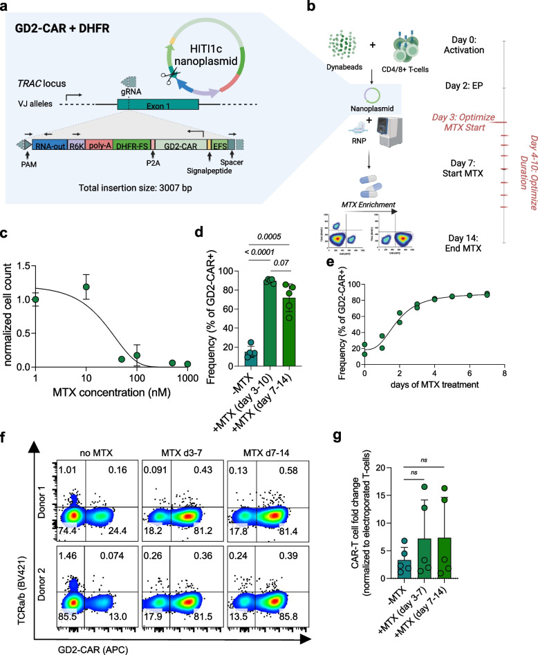
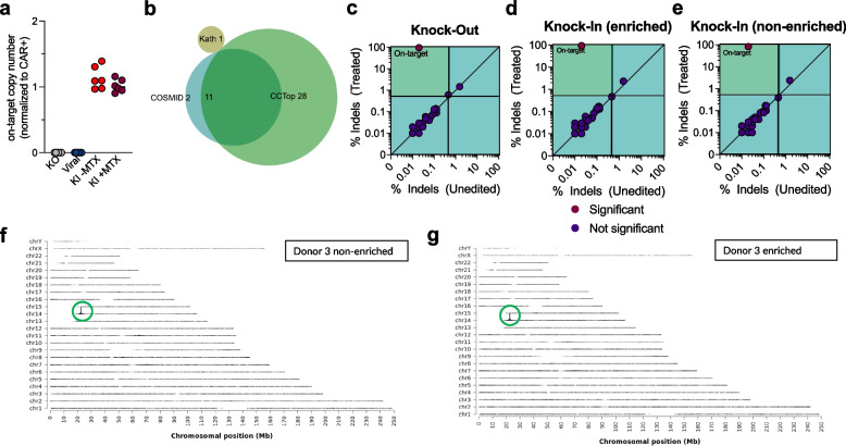
Methods: We applied homology-independent targeted insertion (HITI) or HDR using CRISPR/Cas9 and nanoplasmid DNA to insert an anti-GD2 CAR into the T cell receptor alpha constant (TRAC) locus and compared both targeted insertion strategies in our system. Next, we optimized post-HITI CRISPR EnrichMENT (CEMENT) to seamlessly integrate it into a 14-day process and compared our knock-in with viral transduced anti-GD2 CAR-T cells. Finally, we explored the off-target genomic toxicity of our genomic engineering approach.

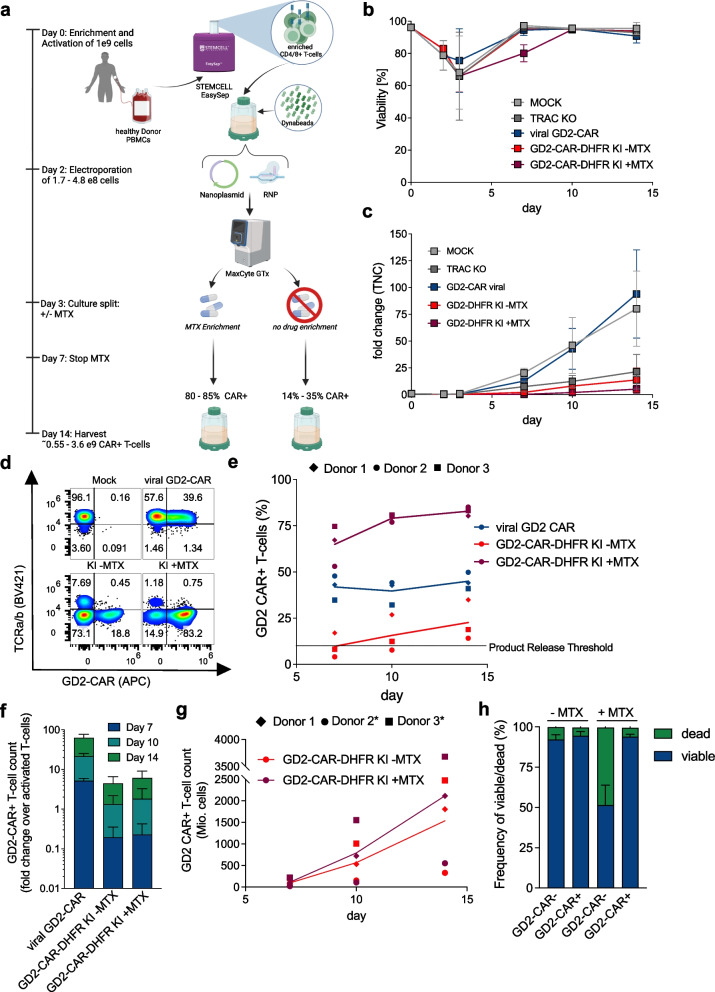
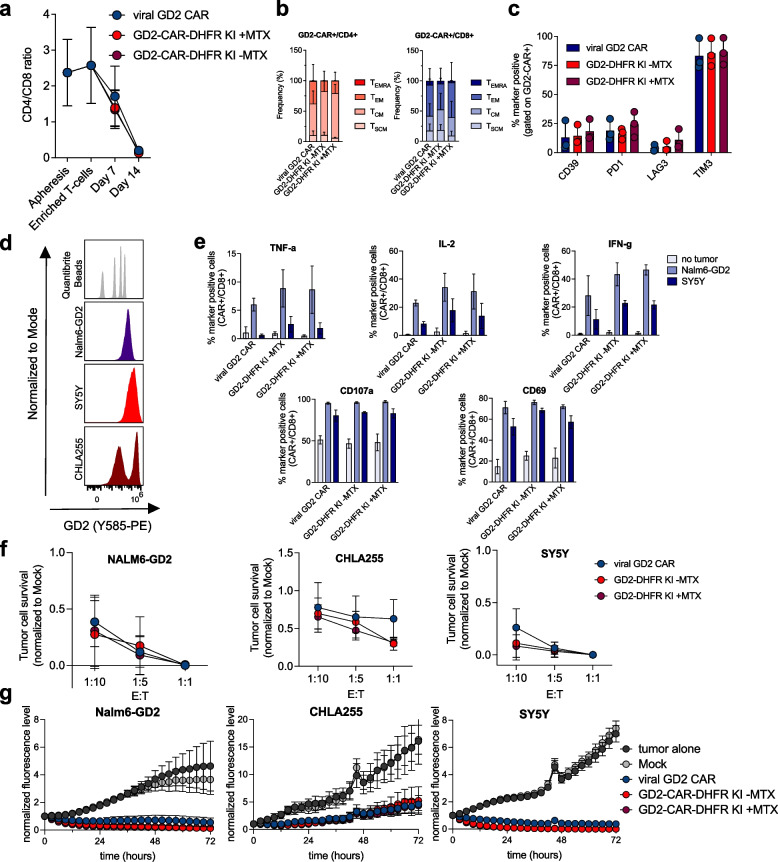
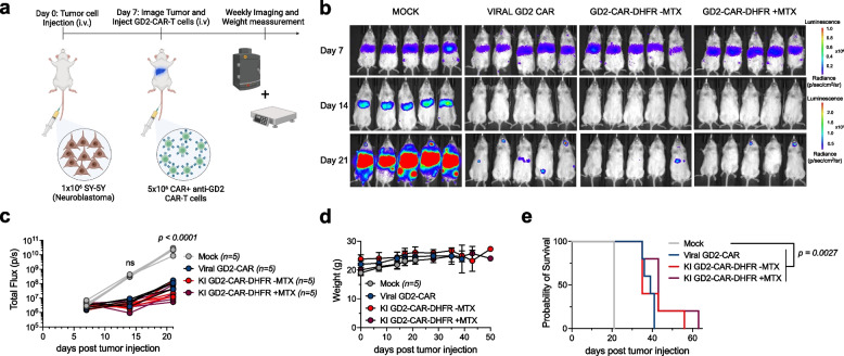
Results: Here, we show that site directed CAR integration utilizing nanoplasmid DNA delivered via HITI provides high cell yields and highly functional cells. CEMENT enriched CAR T cells to approximately 80% purity, resulting in therapeutically relevant dose ranges of 5.5 × 10⁸-3.6 × 10⁹ CAR + T cells. CRISPR knock-in CAR-T cells were functionally comparable with viral transduced anti-GD2 CAR-T cells and did not show any evidence of off-target genomic toxicity.

Conclusions: Our work provides a novel platform to perform guided CAR insertion into primary human T-cells using nanoplasmid DNA and holds the potential to increase access to CAR-T cell therapies.

Keywords: CRISPR/Cas9; GMP; Genomic safety; Homology-independent targeted insertion (HITI); Non-viral CAR-T cell; Targeted insertion.


Conflict of interest statement

H.B.W., C.L.M. and S.A.F. are inventors on a Stanford University provisional patent application pending on Homology-Independent Targeted DNA Insertion in human T cells. A.G.M. has been employed by Maxcyte during this study. G.L.K. is an employee of Integrated DNA Technologies, which sells reagents described in this manuscript. G.L.K owns equity in Danaher Corporation, which is the parent company of Integrated DNA Technologies. Shabnum Patel is an employee of Cargo Therapeutics. E.S. consults for Lepton Pharmaceuticals and Galaria; and holds equity in Lyell Immunopharma. C.L.M. holds patents focused on CAR T cells therapies, is a cofounder and holds equity in Lyell Immunopharma, CARGO Therapeutics and Link Cell Therapies, which are developing CAR-based therapies, and consults for Lyell, CARGO, Link, Apricity, Nektar, Immatics, Mammoth and Ensoma.

Figures

Fig. 1
Fig. 1
Comparison of Homology-Directed Recombination versus Homology-independent-targeted-insertion for targeted knock-in of a GD2-CAR into TRAC. a Schematic overview of workflow for experiments in b-d, and nanoplasmid designs for knock-in templates. b-d head-to-head comparison of constructs HDR2c, HITI2c and HITI1c. 5 × 106 cells were electroporated per condition on day 2 post activation using respective constructs (0.75 µg of nanoplasmid per 1 × 10.6 cells) and analyzed via flow cytometry on day 10 (b, representative donor; c, pooled frequencies) and counted on the same day to assess GD2-CAR-T cell counts (d) (n = 4 independent donors). eh HDR inhibitor induced modulation of GD2-CAR-T cell integration via HITI and HDR. e Schematic related to f–h. f Representative histograms of GD2-CAR expression after CRISPR knock-in with HDR2c or HITI1c templates either left untreated or treated with 1 µM of AZD0156 for 18 h post electroporation. g + h GD2-CAR expression (g) and GD2-CAR-T cell counts (h) normalized to untreated CRISPR knock-in samples after 18 h of treatment with indicated concentrations of AZD0156 (n = 3 independent donors). i-j, CRISPR knock-in of non-activated T cells using HITI1c and HDR2c for knock-in of the GD2-CAR. Indicated are knock-in frequencies (i), Viability (j) and GD2-CAR-T cell yield. Cells were counted and analyzed via flow cytometry on day 10 or 14 (n = 4 independent donors). p values were determined by paired two-tailed t tests. Error bars indicate standard deviation (SD)
Fig. 2
Fig. 2
Optimization of Methotrexate (MTX) based selection of CRISPR knock-in GD2-CAR-DHFR-FS T cells. a GD2-CAR-DHFR-FS nanoplasmid design incorporating a gRNA cut site for linearization of the nanoplasmid and dsDNA break in TRAC with correct transgene insertion indicated. b Experimental layout for optimization of MTX enrichment. MTX treatment from day 7–14 has previously been reported to result in efficient enrichment in viral transduced CAR-DHFR-FS T cells and served as a reference. c Titration of MTX in primary human T cells with efficient killing starting at 50 nM MTX (n = 2 independent donors analyzed in technical duplicates). d Comparison of knock-in frequency determined via flow cytometry on day 14 in GD2-CAR-DHFR-FS T cells either non-enriched, enriched from day 3–10 or from day 7–14 (n = 5 independent donors). e MTX time course after CRISPR knock-in starting on day 3 for up to 7 days with plateaued enrichment after 4 days of treatment. All samples were assessed via flow cytometry on day 14 (n = 2 independent donors). f Quadrant plots indicating TCR-a/b and GD2-CAR surface expression for two representative out of five independent donors non-enriched, enriched from day 3–7 and from day 7–14. Flow cytometry was conducted on day 14. g GD2-CAR-T cell yield at day 14. Fold changes were calculated based on number of electroporated T cells on day 2 (n = 5 independent donors). Experiments in d and g were evaluated for statistical significance by paired, two-tailed t tests. Error bars indicate SD
Fig. 3
Fig. 3
HITI based CRISPR knock-in CAR-T cell manufacturing at clinical scale. a Schematic workflow of leukapheresis processing to manufacture CRISPR KI CAR-T cells at clinical scale. Per Donor 1 × 109 cells were activated and electroporated. Cultures were split up equally and either left untreated or treated with MTX for enrichment. b + c Viability (b) and fold change (c) of respective cultures over time. d Representative quadrant plots (day 14) showing GD2-CAR expression in TRAC positive cells for viral transduced CAR-T cells and TRAC negative cells for GD2 knock-in CAR-T cells. e GD2-CAR frequency over time across all three donors. f expansion of respective GD2-CAR-T cells for different time points normalized to the number of activated T cells. g, Total GD2-CAR-T cell counts for knock-in CAR-T cells (* = Donors with projected numbers after culture split on day 10). h Frequency of viable and dead cells in edited and non-edited T cells after MTX treatment assessed via flow cytometry on day 7. All experiments were conducted with n = 3 independent donors. Error bars indicate SD
Fig. 4
Fig. 4
Knock-in GD2-CAR-T cells do not show phenotypic differences and are not functionally inferior when compared to viral GD2-CAR-T cells. a Changes of CD4/CD8 ratio after processing of leukopaks and over time. b + c Memory marker (b) and exhaustion marker (c) expression of viral vs. GD2 knock-in CAR-T cells as determined via flow cytometry on day 14 (pooled data from n = 3 independent donors). d GD2 antigen levels of co-cultured tumor cell lines. Representative histograms from n = 3 independent experiments. e Intracellular cytokine (TNF-a, IL-2, IFN-g) and activation marker (CD107a, CD69) expression after 6 h of co-culture with respective GD2 expressing tumor cell lines. Shown here is the marker positive cell frequency gated on CD8 + CAR + T cells (pooled data from n = 2 independent donors tested in technical triplicates). f Concentration dependent tumor cell killing after 48 h of co-culture with indicated E:T ratios (pooled data from n = 3 independent donors). g Tumor cell killing over time in GD2 antigen expressing tumor cell lines at a E:T ratio of 1:10 (pooled data from n = 2 independent donors tested in technical triplicates). Error bars indicate SD
Fig. 5
Fig. 5
Knock-in GD2-CAR-T cells efficiently control growth of the SY5Y metastatic Neuroblastoma in vivo model. a Schematic of SY5Y tumor cell injection (1 × 106 on day 0 via tail vein injection) in NSG mice. Confirmed tumor engraftment on day 7 via bioluminescent imaging and consecutive GD2-CAR-T cell treatment on day 7 using 5 × 106 GD2-CAR-T cells applied via tail vein injection followed by weekly imaging of tumor bioluminescence. b Bioluminescent images of treated mice over time with color encoded radiance (p/sec/cm2/sr). c Total flux values (p/s) of all animals over time. Statistical significance was evaluated using two-way ANOVA multiple comparisons along with Dunnett’s test for indicated time points. d Weight of all treated animals over time without relevant changes over baseline. f Kaplan–Meier Survival analysis of treated animals. Statistical significance was evaluated using Mantel-Cox test. Error bars indicate SD
Fig. 6
Fig. 6
Genomic characterization of CRISPR knock-in CAR-T cells. a On-target copy number estimation using ddPCR. Genomic DNA from scale up experiments (n = 3 independent donors) was analyzed in technical duplicates using primers/probe to target Albumin (reference gene, copy number = 2) and primers/probe to target the left insertion site of the GD2-CAR into TRAC. Copy number values were normalized to the frequency of CAR + cells as determined via flow cytometry. b Source of predicted off-target sites. c-e, Quantification of indels in predicted off-target sites and TRAC using CRISPAltRations for samples obtained from Donor 3 of large-scale experiments. Editing was binarily classified using a thresholded Fishers Exact test (p < 0.05) with limitations (> 0.5% indels in treatment; < 0.4% indels in control; > 5,000 reads) for edited samples with (c) knock-out, (d) knock-in without enrichment and (e) knock-in after enrichment (red circle = significant; blue circle = not significant). Indel frequencies were plotted against non-electroporated Mock control samples to highlight pre-existing indels and noise. Quadrants display the limits of classification (bottom left – treatment % indels < 0.5; top right – control % indels > 0.4%; top left – all limitations met and classifiable; bottom right – no limitations met). The top left quadrant contains classifiable events that occur in edited samples and indicates only on-target editing in these samples. f + g Representative insertion site analysis for Donor 3 samples of non-enriched (f) and enriched (g) GD2 knock-in CAR-T cells using TLA. GD2 CAR sequences were inserted into the TRAC locus on chromosome 14 without evidence for off-target insertion

References

    1. Maude SL, Laetsch TW, Buechner J, Rives S, Boyer M, Bittencourt H, et al. Tisagenlecleucel in Children and Young Adults with B-Cell Lymphoblastic Leukemia. N Engl J Med. 2018;378(5):439–448. doi: 10.1056/NEJMoa1709866. - DOI - PMC - PubMed
    1. Wang M, Munoz J, Goy A, Locke FL, Jacobson CA, Hill BT, et al. KTE-X19 CAR T-Cell Therapy in Relapsed or Refractory Mantle-Cell Lymphoma. N Engl J Med. 2020;382(14):1331–1342. doi: 10.1056/NEJMoa1914347. - DOI - PMC - PubMed
    1. Munshi NC, Anderson LD, Shah N, Madduri D, Berdeja J, Lonial S, et al. Idecabtagene Vicleucel in Relapsed and Refractory Multiple Myeloma. N Engl J Med. 2021;384(8):705–716. doi: 10.1056/NEJMoa2024850. - DOI - PubMed
    1. Locke FL, Miklos DB, Jacobson CA, Perales M-A, Kersten M-J, Oluwole OO, et al. Axicabtagene Ciloleucel as Second-Line Therapy for Large B-Cell Lymphoma. N Engl J Med. 2021;386(7):640–654. doi: 10.1056/NEJMoa2116133. - DOI - PubMed
    1. Majzner RG, Ramakrishna S, Yeom KW, Patel S, Chinnasamy H, Schultz LM, et al. GD2-CAR T cell therapy for H3K27M-mutated diffuse midline gliomas. Nature. 2022;603(7903):934–941. doi: 10.1038/s41586-022-04489-4. - DOI - PMC - PubMed

Publication types

LinkOut - more resources