. 2023 Jun 26;25(1):75.
doi: 10.1186/s13058-023-01663-y.

Transcriptionally regulated miR-26a-5p may act as BRCAness in Triple-Negative Breast Cancer

Affiliations

Transcriptionally regulated miR-26a-5p may act as BRCAness in Triple-Negative Breast Cancer

Yue Zhang et al. Breast Cancer Res. .

Abstract

Background: DNA damage and DNA damage repair (DDR) are important therapeutic targets for triple-negative breast cancer (TNBC), a subtype with limited chemotherapy efficiency and poor outcome. However, the role of microRNAs in the therapy is emerging. In this study, we explored whether miR-26a-5p could act as BRCAness and enhance chemotherapy sensitivity in TNBC.

Methods: Quantitative reverse transcription polymerase chain reaction (RT-qPCR) was used to detect the expression of miR-26a-5p in breast cancer tissues and cell lines. CCK-8 was used to measure drug sensitivity in concentration gradient and time gradient. Comet assay was used to detect DNA damage. Flow cytometry was performed to examine apoptosis. Moreover, we used western blot and immunofluorescence to detect biomarkers. Luciferase reporter assay was performed to verify the combination of miR-26a-5p and 3'UTR of target gene. Hormone deprivation and stimulation assay were used to validate the effect of hormone receptors on the expression of miR-26a-5p. Chromatin immunoprecipitation (ChIP) assays were used to verify the binding sites of ER-a or PR with the promoter of miR-26a-5p. Animal experiments were performed to the effect of miR-26a-5p on Cisplatin treatment.

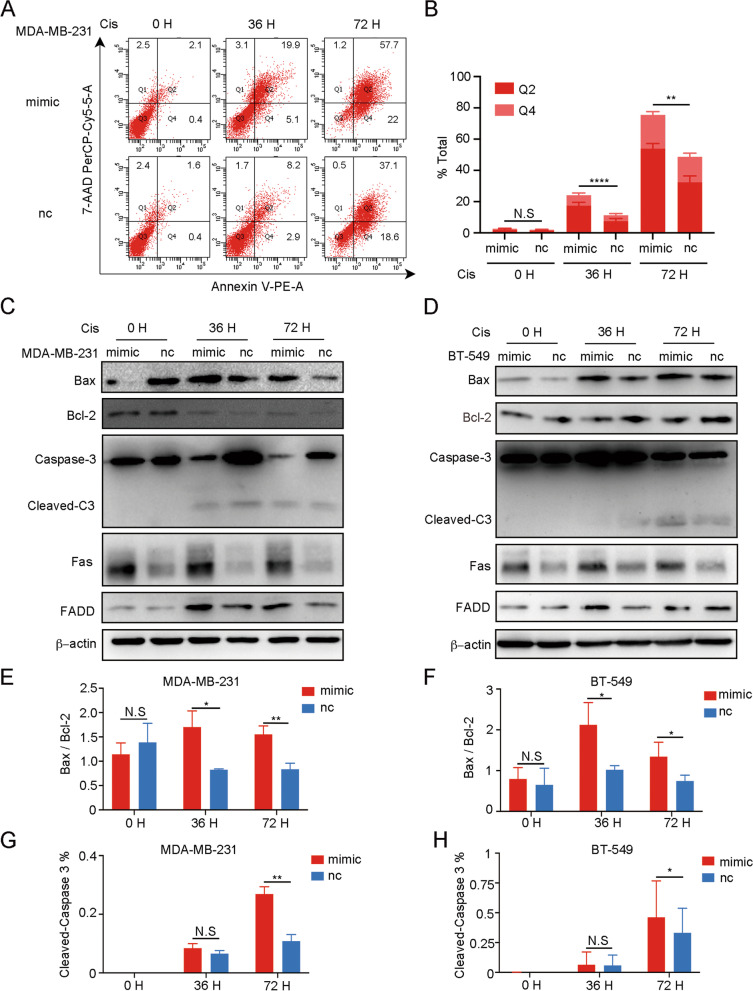
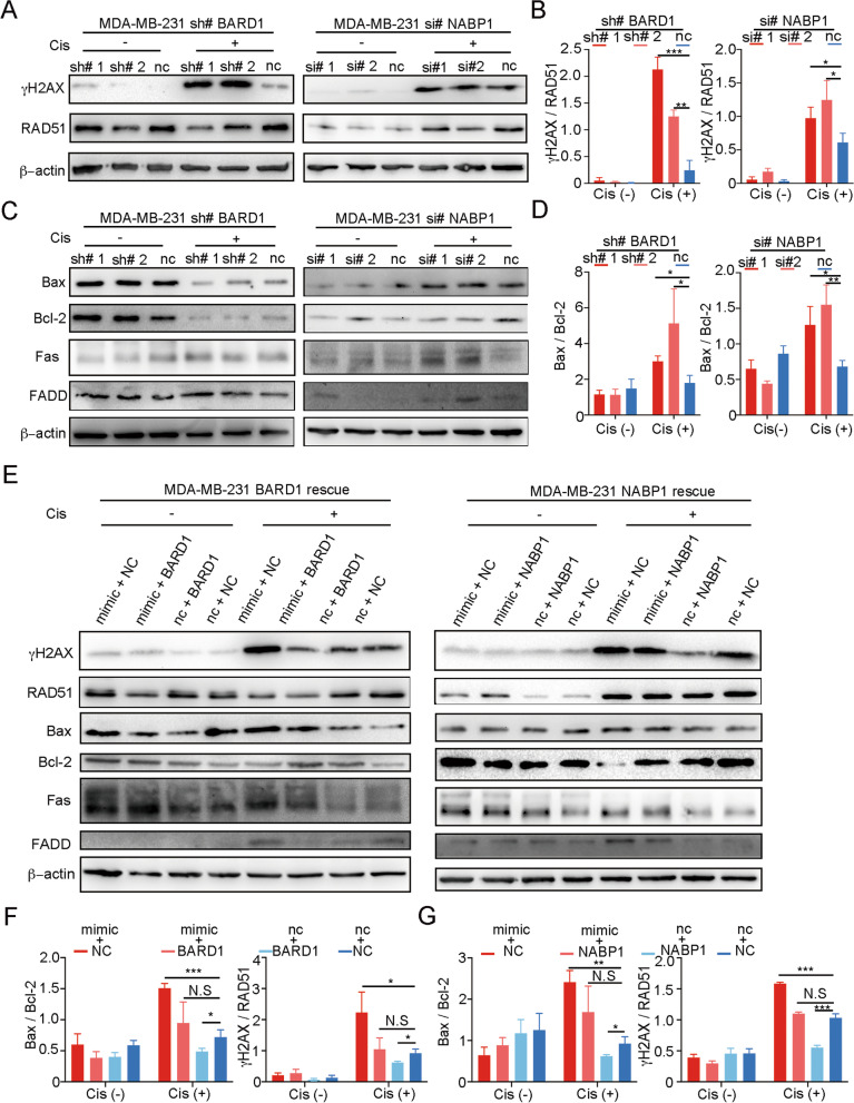
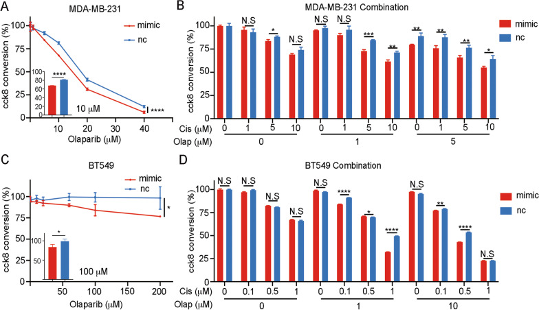
Results: The expression of miR-26a-5p was significantly downregulated in TNBC. Overexpressing miR-26a-5p enhanced the Cisplatin-induced DNA damage and following apoptosis. Interestingly, miR-26a-5p promoted the expression of Fas without Cisplatin stimulating. It suggested that miR-26a-5p provided a hypersensitivity state of death receptor apoptosis and promoted the Cisplatin sensitivity of TNBC cells in vitro and in vivo. Besides, miR-26a-5p negatively regulated the expression of BARD1 and NABP1 and resulted in homologous recombination repair defect (HRD). Notably, overexpressing miR-26a-5p not only facilitated the Olaparib sensitivity of TNBC cells but also the combination of Cisplatin and Olaparib. Furthermore, hormone receptors functioned as transcription factors in the expression of miR-26a-5p, which explained the reasons that miR-26a-5p expressed lowest in TNBC.

Conclusions: Taken together, we reveal the important role of miR-26a-5p in Cisplatin sensitivity and highlight its new mechanism in DNA damage and synthetic lethal.

Keywords: Cisplatin; DNA damage; HRD; Synthetic lethal; TNBC; miR-26a-5p.


Conflict of interest statement

The authors declare that they have no competing interests.

Figures

Fig. 1
Fig. 1
miR-26a-5p is downregulated in TNBC. A Downregulated miRNAs in breast cancer tissue especially in TNBC were obtained from TCGA-BRCA database. B Heatmap of different downregulated miRNAs expression between TNBC and normal breast tissue from TCGA-BRCA database. C MirPath v.3 GO Reverse Search was used to find DNA damage-related miRNAs. Four candidate miRNAs were chosen from overlapping the above four gene sets and we chose miR-26a-5p as a preferred target. D The expression of miR-26a-5p in breast cancer (red bar) was downregulated compared with normal tissues (blue bar) from TCGA-BRCA database. Its expression in TNBC (red bar) was downregulated compared with NTNBC (blue bar). E The expression of miR-26a-5p in breast cancer tissues (red bar) and matched normal tissues (blue bar) was measured by RT-qPCR. Its expression in TNBC (red bar) was downregulated compared with NTNBC (blue bar) in clinical tissues. F The expression of miR-26a-5p in breast cancer cell lines and human normal breast epithelial cell line MCF10A was measured by RT-qPCR. ***P < 0.001, ****P < 0.0001
Fig. 2
Fig. 2
miR-26a-5p upregulation promotes the sensitivity of Cisplatin and DNA damage in TNBC cell lines. A, B Cisplatin-insensitive (MDA-MB-231) and Cisplatin-sensitive (BT549) TNBC cell lines were chosen to transfect miR-26a-5p mimics and normal control. Different concentrations of Cisplatin were diluted with medium containing 1% FBS to incubate cell lines for 72 h. CCK8 was used to detect cytotoxicity. C–E Comet assay was used to identify the level of DNA damage after Cisplatin treatment to MDA-MB-231 and BT549 cells overexpressing miR-26a-5p, respectively, in 20 µM and 2 µM. F CCK8 was used to detect the live cell count at different time points after a special concentration of Cisplatin treatment (MDA-MB-231, 20 µM and BT549, 2 µM). G Western blot was used to measure the expression of DNA damage mark, γH2AX, in MDA-MB-231 and BT549 cells overexpressing miR-26a-5p at different time points after Cisplatin treatment. H, I Immunofluorescence was adopted to detect γH2AX expression change in the MDA-MB-231 cells exposed to Cisplatin for 6 h. **P < 0.01, ***P < 0.001, ****P < 0.0001 vs. control. N.S. not significant. n = 3
Fig. 3
Fig. 3
miR-26a-5p upregulation promotes apoptosis caused by Cisplatin-induced DNA damage. A, B Flow cytometry was used to detect early apoptosis and late apoptosis in the MDA-MB-231 cells overexpressing miR-26a-5p after Cisplatin exposure at different time points. C, D The expression of apoptosis markers in MDA-MB-231 and BT549 cells overexpressing miR-26a-5p after Cisplatin exposure was measured by Western blot. E–H The gray values were detected by Image J and then got the gray values ratio. **P < 0.01, ***P < 0.001, ****P < 0.0001 vs. control. N.S not significant. n = 3
Fig. 4
Fig. 4
miR-26a-5p directly targets the 3’UTR of BARD1 and NABP1. A MicroT-CDS software was used to predict the downstream gene of miR-26a-5p. DDR gene sets were selected from GSEA randomly and then overlapped the three gene sets and got three potential target genes, HMGA1, NABP1 and BARD1. B BARD1 and NABP1 proteins were restrained after overexpressing miR-26a-5p in MDA-MB-231 and BT549 cells measured by Western blot. C, D TargetScan website was used to predict binding sites of miR-26a-5p and BARD1 mRNA. Dual-luciferase reporter assay was performed to confirm binding sites in 293T cells. E, F TargetScan website was used to predict binding sites of miR-26a-5p and NABP1 mRNA. Dual-luciferase reporter assay was performed to confirm binding sites in 293T cells. G–J CCK8 was used to measured cytotoxicity of Cisplatin after knockdown BARD1 in shRNA and NABP1 in siRNA in TNBC cells, respectively. *P < 0.05, **P < 0.01, ***P < 0.001, ****P < 0.0001 vs. control. N.S not significant. n = 3
Fig. 5
Fig. 5
miR-26a-5p promotes Cisplatin-induced cell death via BARD1 and NABP1. AD MDA-MB-231 cells were knockdown BARD1 in shRNA and NABP1 in siRNA, respectively. DNA damage marker (γH2AX), DDR marker (RAD51) and apoptosis markers were detected without or with 20 µM Cisplatin stimulating by Western blot. E–G BARD1 or NABP1 was transfected into miR-26a-5p-overexpressing MDA-MB-231 cells. Western blot was performed to evaluate the expression of the markers with or without Cisplatin stimulating. The gray values were detected by Image J and then got the gray values ratio. *P < 0.05, **P < 0.01, ***P < 0.001, ****P < 0.0001 vs. control. N.S not significant. n = 3
Fig. 6
Fig. 6
In vivo effect of miRNA-26a-5p on the Cisplatin sensitivity. A, B The indicated cells were implanted orthotopically into the second mammary fat pad (i.m.f.p.) of female BALB/C nude mice. The volumes of subcutaneous tumors in the indicated groups (n = 3) were recorded. C RTV was introduced to illustrate Cisplatin effect on Cisplatin sensitivity in vivo. D, E T/C% was introduced to evaluate drug efficacy in vivo. F–H Representative images of IHC staining and the relative scores (n = 12) of the indicated markers in subcutaneous tumors of mice. *P < 0.05, **P < 0.01, ***P < 0.001, ****P < 0.0001 versus control. N.S not significant
Fig. 7
Fig. 7
miR-26a-5p upregulation promotes the sensitivity of Olaparib and drug combination. A MDA-MB-231 cells were transfected with miR-26a-5p mimics and normal control. Different concentrations of Olaparib were diluted with medium containing 1% FBS to incubate cell lines for 72 h. CCK8 was used to detect cytotoxicity. B CCK8 was used to detect cytotoxicity of the drug combination of Cisplatin and Olaparib in MDA-MB-231 overexpressing miR-26a-5p and normal control. C, D CCK8 was used to detect the indicated drug effect on BT549 cells. *P < 0.05, **P < 0.01, ***P < 0.001, ****P < 0.0001 vs. control. N.S not significant. n = 3
Fig. 8
Fig. 8
miR-26a-5p is a transcriptional downstream target of ER-a and PR. A bc-GenExMiner was used to analyze the expression level of miR-26a-5p which was positively correlated with ER-a status and PR status, not HER2 receptor status. B After hormone deprivation for 48 h, MCF-7 cells were stimulated with β-Estradiol (E2). RT-qPCR was used to monitor the expression of miR-26a-5p in different concentration or at different time points. C After hormone deprivation for 48 h, MCF-7 cells were stimulated with Etonogestrel (ETO). RT-qPCR was used to monitor the expression of miR-26a-5p in different concentration or at different time points. D, E Overexpressed ER-a or PR plasmid were transfected into MDA-MB-231 cells. RT-qPCR was used to detect the expression of miR-26a-5p in normal culture conditions without or with extra hormone stimulating. F JASPAR and hTFtarget databases were used to predict the potential binding sites of ER-a and PR with the promoter of miR-26a-5p. G, H ChIP assay was used to verify the binding sites. *P < 0.05, **P < 0.01, ***P < 0.001, ****P < 0.0001 versus control. n = 3

References

    1. Siegel RL, Miller KD, Fuchs HE, Jemal A. Cancer statistics, 2022. CA Cancer J Clin. 2022;72(1):7–33. doi: 10.3322/caac.21708. - DOI - PubMed
    1. McDonald ES, Clark AS, Tchou J, Zhang P, Freedman GM. Clinical Diagnosis and Management of Breast Cancer. J Nucl Med. 2016;57(Suppl 1):9S–16S. doi: 10.2967/jnumed.115.157834. - DOI - PubMed
    1. Prat A, Lluch A, Albanell J, Barry WT, Fan C, Chacon JI, et al. Predicting response and survival in chemotherapy-treated triple-negative breast cancer. Br J Cancer. 2014;111(8):1532–1541. doi: 10.1038/bjc.2014.444. - DOI - PMC - PubMed
    1. O'Hara SP, Mott JL, Splinter PL, Gores GJ, LaRusso NF. MicroRNAs: key modulators of posttranscriptional gene expression. Gastroenterology. 2009;136(1):17–25. doi: 10.1053/j.gastro.2008.11.028. - DOI - PMC - PubMed
    1. Cheng C, Guo L, Ma Y, Wang Z, Fan X, Shan Z. Up-regulation of miR-26a-5p inhibits E2F7 to regulate the progression of renal carcinoma cells. Cancer Manag Res. 2020;12:11723–11733. doi: 10.2147/CMAR.S271710. - DOI - PMC - PubMed

Publication types