. 2023 Nov;19(11):5307-5315.
doi: 10.1002/alz.13352. Epub 2023 Jun 27.

Hippocampal sclerosis of aging at post-mortem is evident on MRI more than a decade prior

Affiliations

Hippocampal sclerosis of aging at post-mortem is evident on MRI more than a decade prior

Diana Ortega-Cruz et al. Alzheimers Dement. 2023 Nov.

Abstract

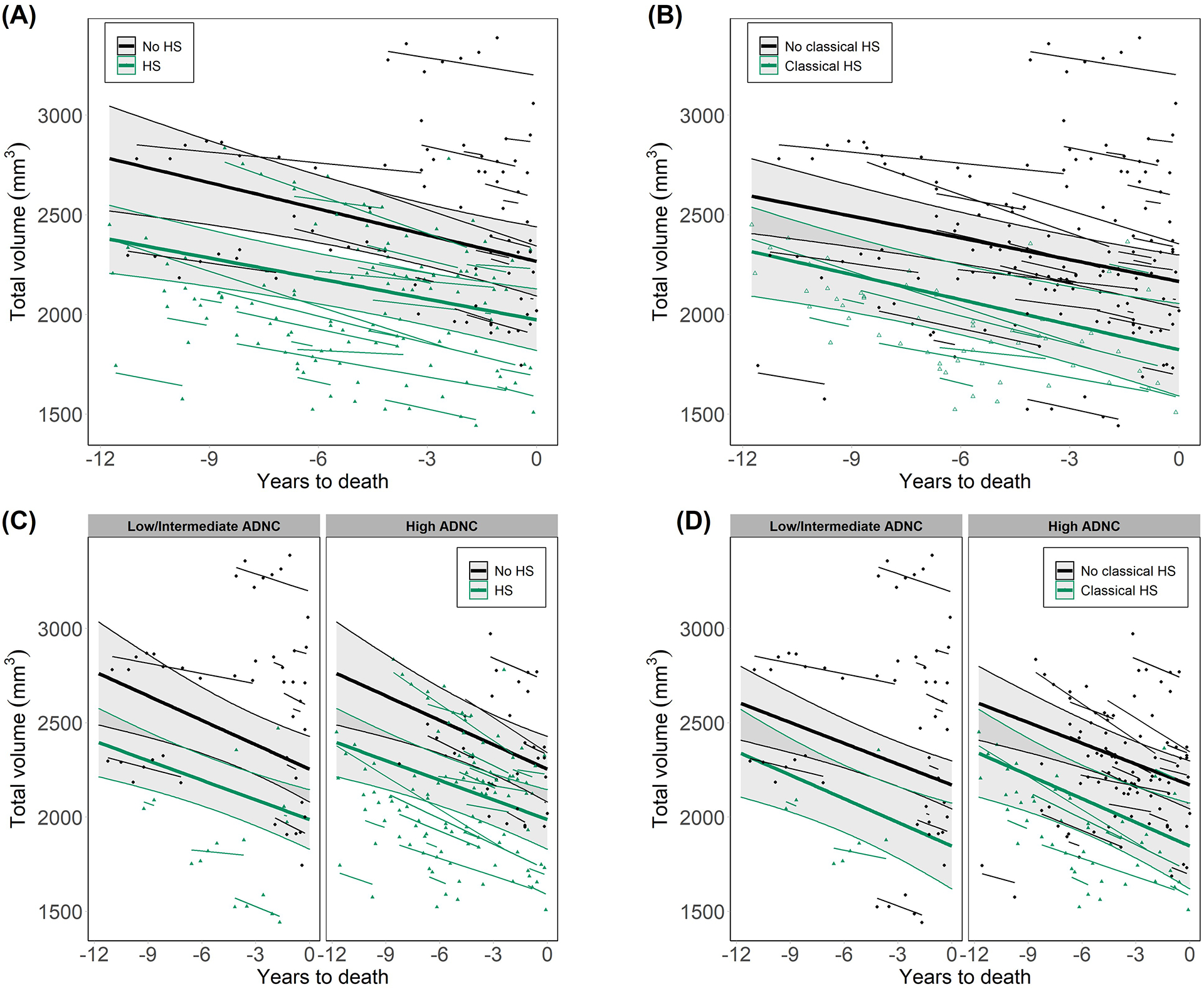
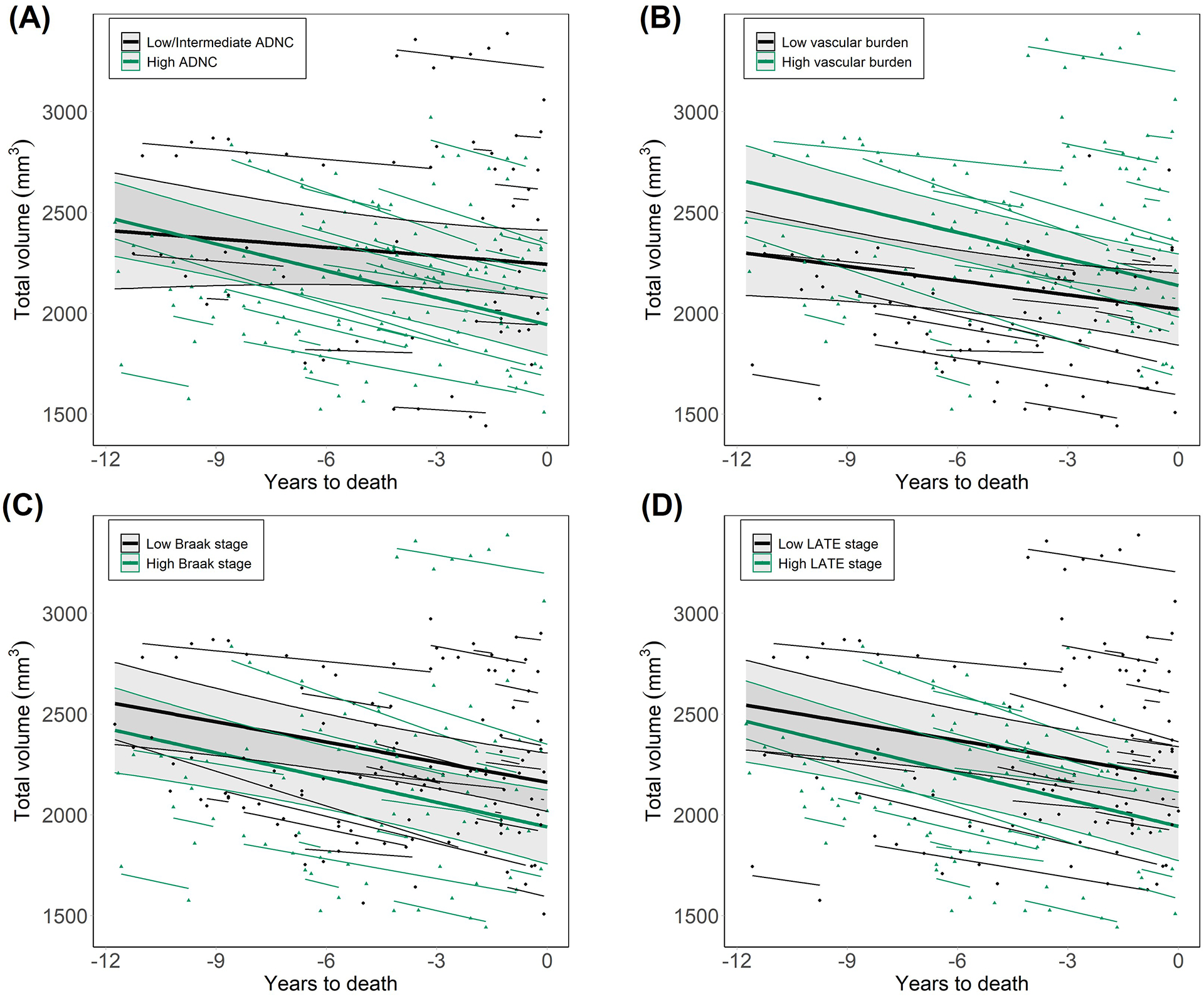
Introduction: Hippocampal sclerosis of aging (HS) is an important component of combined dementia neuropathology. However, the temporal evolution of its histologically-defined features is unknown. We investigated pre-mortem longitudinal hippocampal atrophy associated with HS, as well as with other dementia-associated pathologies.

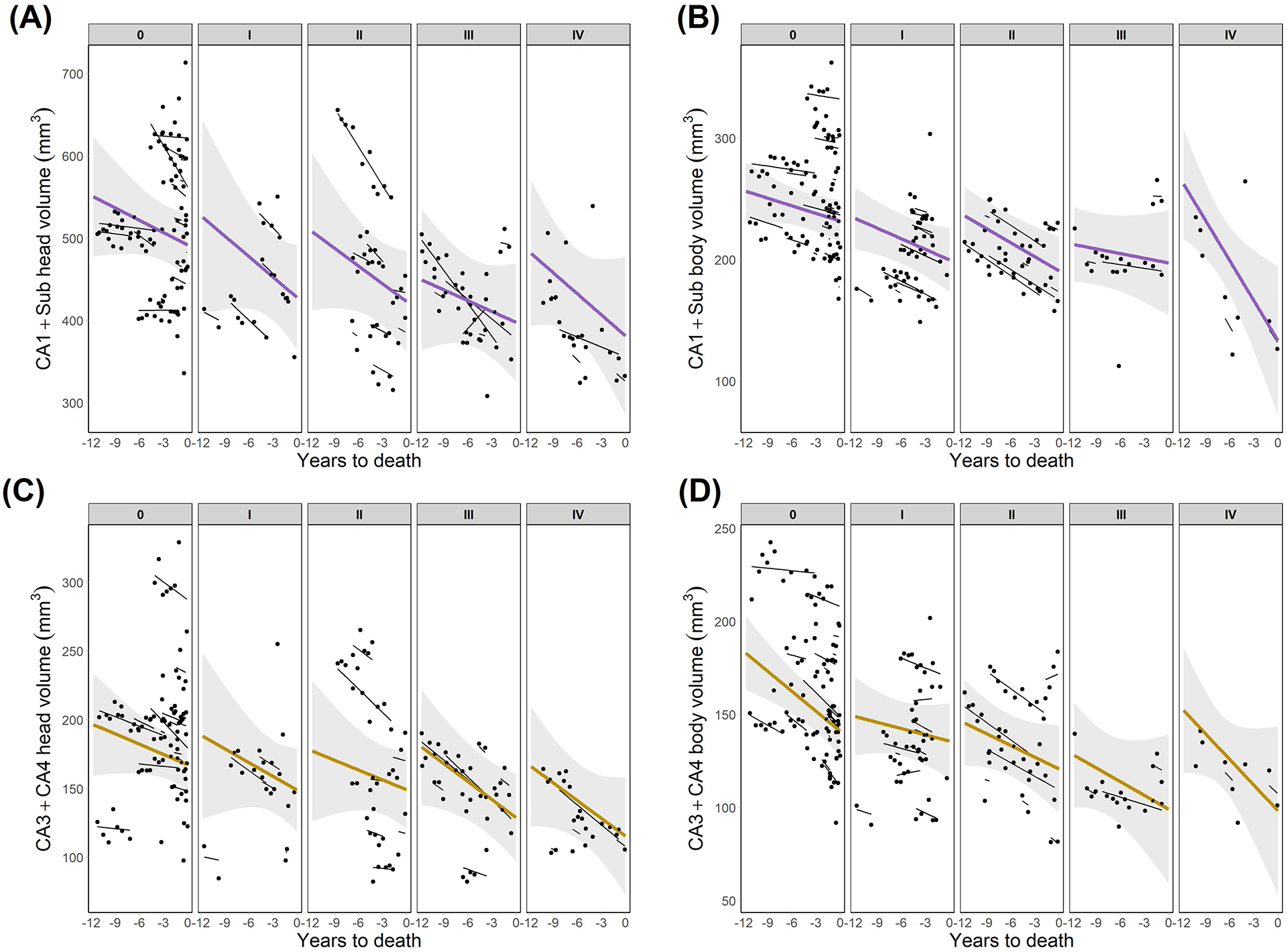
Methods: We analyzed hippocampal volumes from magnetic resonance imaging (MRI) segmentations in 64 dementia patients with longitudinal MRI follow-up and post-mortem neuropathological evaluation, including HS assessment in the hippocampal head and body.

Results: Significant HS-associated hippocampal volume changes were observed throughout the evaluated timespan, up to 11.75 years before death. These changes were independent of age and Alzheimer's disease (AD) neuropathology and were driven specifically by CA1 and subiculum atrophy. AD pathology, but not HS, was associated significantly with the rate of hippocampal atrophy.

Discussion: HS-associated volume changes are detectable on MRI earlier than 10 years before death. Based on these findings, volumetric cutoffs could be derived for in vivo differentiation between HS and AD.

Highlights: Hippocampal atrophy was found in HS+ patients earlier than 10 years before death. These early pre-mortem changes were driven by reduced CA1 and subiculum volumes. Rates of hippocampus and subfield volume decline were independent of HS. In contrast, steeper atrophy rates were associated with AD pathology burden. Differentiation between AD and HS could be facilitated based on these MRI findings.

Keywords: atrophy; dementia; hippocampal sclerosis of aging; hippocampus; longitudinal; magnetic resonance imaging; neuropathology.


Conflict of interest statement

Conflicts

Declarations of interest: none

Figures

Figure 1.
Figure 1.. Longitudinal progression of hippocampal volumes as a function of HS.
(A) HS groups defined as a function of HS staging including early and advanced stages (HS+ for those at stage>0). (B) HS groups determined by the classical definition of the pathology, by which only advanced cases with severe cell loss are included within the HS+ group. (C) HS groups defined as a function of HS staging, compared within groups of high and low/intermediate ADNC. After including ADNC grouping in the statistical model, the effect of HS was still significant (F=30.7, p=.015), with lower volumes in HS+ compared to HS− patients at 11.75 years before death. (D) HS groups determined by the classical definition of the pathology, compared within ADNC burden groups, with the effect of HS remaining significant after including this variable (F=26.1, p=.006). ADNC: Alzheimer’s disease neuropathological change. HS: hippocampal sclerosis of aging.
Figure 2.
Figure 2.. Longitudinal progression of hippocampal subfields volumes as a function of HS staging.
(A) Volumes of CA1+subiculum in the head of the hippocampus. (B) Volumes of CA1+subiculum in the hippocampal body. (C) CA3+CA4 volumes in the head of the hippocampus. (D) CA3+CA4 volumes in the hippocampal body. CA1+Sub: CA1+subiculum.
Figure 3.
Figure 3.. Longitudinal volumes of the whole hippocampus as a function of other neuropathologies in dementia.
(A) Differences between groups of low/intermediate and high probability of ADNC. Significant volume differences between these groups were found (F=13.4, p=0.004), as well as significantly different slopes (p=.016, estimated slopes (mm3/year): −14 for low/intermediate, −44.5 for high ADNC). To evaluate the effects of pathologies considered in B, C and D, the contribution of ADNC was taken into account by adding it as a covariate in these models. (B) Groups of low and high vascular pathology burden, following the evaluation proposed by Deramecourt et al., showed no significant between-group volume differences (F=7.8, p=.294). (C) Groups of low and high Lewy body pathology burden, as a function of Braak staging for α-synuclein. Differences between these groups showed a tendency to significance (F=4.7, p=.084). (D) Comparisons between groups of low and high TDP-43 pathology burden (F=14.6, p=0.022), evaluated through LATE staging. ADNC: Alzheimer’s disease neuropathological change. LATE: limbic age-related TDP-43 encephalopathy.

Update of

Similar articles

Cited by

References

    1. Amador-Ortiz C, Ahmed Z, Zehr C, Dickson D. Hippocampal sclerosis dementia differs from hippocampal sclerosis in frontal lobe degeneration. Acta Neuropathol. 2007;113(3):245–252. doi:10.1007/s00401-006-0183-4 - DOI - PMC - PubMed
    1. Nelson PT, Fardo DW, Van Eldik LJ, et al. Hippocampal Sclerosis of Aging, a Prevalent and High-Morbidity Brain Disease. Acta Neuropathol. 2013;126(2):161–177. doi:10.1007/s00401-013-1154-1 - DOI - PMC - PubMed
    1. Dickson DW, Davies P, Bevona C, et al. Hippocampal sclerosis: a common pathological feature of dementia in very old (≥80 years of age) humans. Acta Neuropathol. 1994;88(3):212–221. doi:10.1007/BF00293396 - DOI - PubMed
    1. Nelson PT, Schmitt FA, Lin Y, et al. Hippocampal sclerosis in advanced age: Clinical and pathological features. Brain. 2011;134(5):1506–1518. doi:10.1093/brain/awr053 - DOI - PMC - PubMed
    1. Aoki N, Murray ME, Ogaki K, et al. Hippocampal sclerosis in Lewy body disease is a TDP-43 proteinopathy similar to FTLD-TDP Type A. Acta Neuropathol. 2015;129(1):53–64. doi:10.1007/s00401-014-1358-z - DOI - PMC - PubMed