Review
. 2021 Jun 15:4:25152564211022515.
doi: 10.1177/25152564211022515. eCollection 2021 Jan-Dec.

Mutant VAPB: Culprit or Innocent Bystander of Amyotrophic Lateral Sclerosis?

Affiliations
Review

Mutant VAPB: Culprit or Innocent Bystander of Amyotrophic Lateral Sclerosis?

Nica Borgese et al. Contact (Thousand Oaks). .

Abstract

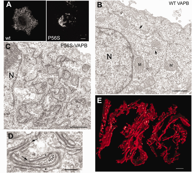
Nearly twenty years ago a mutation in the VAPB gene, resulting in a proline to serine substitution (p.P56S), was identified as the cause of a rare, slowly progressing, familial form of the motor neuron degenerative disease Amyotrophic Lateral Sclerosis (ALS). Since then, progress in unravelling the mechanistic basis of this mutation has proceeded in parallel with research on the VAP proteins and on their role in establishing membrane contact sites between the ER and other organelles. Analysis of the literature on cellular and animal models reviewed here supports the conclusion that P56S-VAPB, which is aggregation-prone, non-functional and unstable, is expressed at levels that are insufficient to support toxic gain-of-function or dominant negative effects within motor neurons. Instead, insufficient levels of the product of the single wild-type allele appear to be required for pathological effects, and may be the main driver of the disease. In light of the multiple interactions of the VAP proteins, we address the consequences of specific VAPB depletion and highlight various affected processes that could contribute to motor neuron degeneration. In the future, distinction of specific roles of each of the two VAP paralogues should help to further elucidate the basis of p.P56S familial ALS, as well as of other more common forms of the disease.

Keywords: endoplasmic reticulum (ER); intracellular inclusions; membrane contact sites; motor neuron; neurodegeneration; phosphoinositide.


Conflict of interest statement

The author(s) declared no potential conflicts of interest with respect to the research, authorship, and/or publication of this article.

Figures

Figure 1.
Figure 1.
The VAP proteins. (a): Domain organization of VAPA and B. The orientation of the two proteins with respect to the ER membrane and the percent of shared sequence between the two human isoforms are indicated. (b) Multiple sequence alignment (https://www.ebi.ac.uk/Tools/msa/clustalo/) of MSP of mammalian, invertebrate and fungal VAP homologues. Note the remarkable conservation of residues (enclosed in red boxes) at the binding interface with an FFAT- containing peptide (Shi et al., 2010). The asterisks indicate the Thr and Pro residues substituted in the p.T46I and p.P56S mutants. The green arrows indicate the D1 and D2 β strands (nomenclature from (Kaiser et al., 2005)) bridged by an S-shaped loop centred on P56. (c) Images are slightly modified from (Furuita et al., 2010). Left: ribbon representation of the VAPA MSP (nomenclature of the β strands according to (Kaiser et al., 2005)) in complex with an FFAT-containing peptide derived from OSBP, shown in orange as a stick model. The position of Pro56 is highlighted in cyan. Right: Details of the interaction between VAPA-MSP and OSBP FFAT peptide. Residue numbers of OSBP peptide, shown as stick model, are indicated in italics. The surface of VAPA MSP is represented with acidic (red), basic (blue), and hydrophobic (yellow) residues. Thr46 is boxed.
Figure 2.
Figure 2.
Summary of the functions of the VAP proteins. See text for further explanation.
Figure 3.
Figure 3.
Possible mechanisms of dominance of the p.P56S mutation. Top: heterozygous individuals produce P56S-VAPB (red) and the wt protein (green). Left: toxic gain of function of the mutant: P56S-VAPB aggregates, or smaller oligomeric assemblies, could be harmful to the cell by sequestering proteins involved in proteostasis or other basic cellular processes, or by unphysiologically trapping normal binding partners (sequestered proteins are represented by blue circles). Right: Loss of function of the wt allele: this could occur by negative dominance, i.e., inactivation of the wt protein by sequestration into mutant aggregates; alternatively, P56S-VAPB inclusions could be without effect and easily degraded (red dots), but the amount of protein generated from the single wt allele would be insufficient to sustain normal function (haploinsufficiency).
Figure 4.
Figure 4.
Characterisation of inclusions formed by P56S-VAPB. (A) Immunofluorescence of HeLa cell expressing wt or P56S-VAPB. (B and C) Transmission electron micrographs of HeLa cells transiently transfected with wt (B) or P56S-VAPB (C). N, nucleus, M, mitochondrion; the arrows in (B) indicate normal ER cisternae. (D) high magnification view of altered ER induced by P56S-VAPB. The asterisks mark the ER lumen and the arrows indicate the continuity of the electron dense layer between the cisternae with the surrounding cytosol. The five small arrowheads indicate membrane-attached ribosomes. (E) Tomographic 3D reconstruction of P56S-VAPB inclusion in transfected HeLa cell, confirming the presence of ER cisternae in the structures. Panels (A) to (D): modified from (Fasana et al., 2010), panel (E) is from (Papiani et al., 2012). Scale bars: (A), 5 μm; (B) and (), 1 μm; (D) and (E), 200 nm.

Similar articles

Cited by

References

    1. Al-Chalabi A, van den Berg LH, Veldink J. (2017). Gene discovery in amyotrophic lateral sclerosis: implications for clinical management. Nat Rev Neurol 13, 96–104. doi: 10.1038/nrneurol.2016.182 - PubMed
    1. Aliaga L, Lai C, Yu J, Chub N, Shim H, Sun L, Xie C, Yang WJ, Lin X, O'Donovan MJ, Cai H. (2013). Amyotrophic lateral sclerosis-related VAPB P56S mutation differentially affects the function and survival of corticospinal and spinal motor neurons. Human Mol Genet 22, 4293–4305. doi: 10.1093/hmg/ddt279 - PMC - PubMed
    1. Alpy F, Rousseau A, Schwab Y, Legueux F, Stoll I, Wendling C, Spiegelhalter C, Kessler P, Mathelin C, Rio MC, et al.. (2013). STARD3 or STARD3NL and VAP form a novel molecular tether between late endosomes and the ER. J Cell Sci 126, 5500–5512. doi: 10.1242/jcs.139295 - PubMed
    1. Anagnostou G, Akbar MT, Paul P, Angelinetta C, Steiner TJ, de Belleroche J. (2010). Vesicle associated membrane protein B (VAPB) is decreased in ALS spinal cord. Neurobiol Aging 31, 969–985. doi: 10.1016/j.neurobiolaging.2008.07.005 - PubMed
    1. Antonny B, Bigay J, Mesmin B. (2018). The oxysterol-binding protein cycle: burning off PI(4)P to transport cholesterol. Annu Rev Biochem 87, 809–837. doi: 10.1146/annurev-biochem-061516-044924 - PubMed

LinkOut - more resources