Case Reports
. 2023 May 26;15(5):e39539.
doi: 10.7759/cureus.39539. eCollection 2023 May.

Delayed Sub-axial Fracture Dislocation Surgical Management: Technical Notes and Review of the Literature

Affiliations
Case Reports

Delayed Sub-axial Fracture Dislocation Surgical Management: Technical Notes and Review of the Literature

Fahad Alhelal et al. Cureus. .

Abstract

The surgical treatment of delayed, unstable sub-axial cervical spine injuries is challenging. Multiple treatment regimens have been described in the literature, although there is no consensus regarding the best treatment approach. This report presents a 35-year-old obese woman who experienced a delayed sub-axial fracture-dislocation following a motor vehicle accident (MVA) and was successfully managed after three weeks via pre-operative traction followed by a novel single-surgery, single-approach technique with pedicle screws and tension-band wiring as a reduction method. A 35-year-old obese woman with a body mass index (BMI) of 30.1 sustained a frontal impact MVA and suffered from complete quadriplegia below C5 (American Spinal Cord Association Injury A) three weeks prior to presentation. She was intubated and presented with a Glasgow Coma Scale score of 11/15. Trauma computed tomography (CT) showed an isolated spine injury. Moreover, whole-spine CT showed an isolated cervical spine injury involving a basin tip fracture, a comminuted C1 arch fracture, a C2 fracture, and a C6-C7 fracture-dislocation. In addition, magnetic resonance imaging revealed cord contusion at the same level, with C1-C2 left atlantoaxial joint instability. Neck magnetic resonance angiograms and carotid CT angiograms showed left vertebral artery attenuation. She was admitted to the intensive care unit and taken for C6-C7 reduction and instrumentation using only a posterior approach after medical optimization and the application of sufficient traction. Delayed cervical spine fracture-dislocation imposes a challenge for surgical reduction. However, a proper reduction can be achieved through a sufficient duration of pre-operative traction and an isolated anterior or posterior approach.

Keywords: cervical vertebrae; delayed sub-axial injuries; fracture dislocation; quadriplegia; vertebral artery injury.


Conflict of interest statement

The authors have declared that no competing interests exist.

Figures

Figure 1
Figure 1. CT scan of C6-7 fracture dislocation
(A) Cervical spine CT scan sagittal cuts demonstrating a total fracture dislocation of C6 over C7; (B) cervical spine CT scan axial cuts demonstrating the corresponding level of C6–C7.
Figure 2
Figure 2. CT scan of C5-6 fracture involvement
(A) Cervical spine CT scan sagittal cuts demonstrating a comminuted fracture of C5 transverse process, bilateral pedicles, and spinous process; (B) cervical spine CT scan axial cuts demonstrating the corresponding levels of C5 and C6–C7.
Figure 3
Figure 3. CT scan depicting C2 fracture
(A,C) Cervical spine CT scan sagittal and coronal cuts demonstrating a fracture dissecting through the base of C2 and pars interarticularis and bilateral transverse foramina of C2. (B,D) Axial cervical spine CT cuts of the corresponding level of C1–C2 vertebral body.
Figure 4
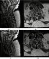
Figure 4. MRI of C6-C7 fracture dislocation
(A,C): Sagittal spine MRI demonstrating a total fracture dislocation of C6–C7 with marked cord contusion. (B,D) Axial spine MRI of the corresponding level of C6–C7 vertebral body.
Figure 5
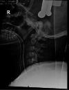
Figure 5. Intra-op lateral cervical X-ray
Lateral cervical spine X-ray showing Gardner–Wells traction.
Figure 6
Figure 6. X-rays of final operative reduction and fixation
(A) Lateral cervical spine X-ray demonstrating final fixation from C3–T2; (B) AP spine X-ray demonstrating the corresponding level.
Figure 7
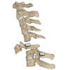
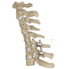
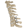
Figure 7. C6-7 transpedicular screw fixation
Illustration of transpedicular screws fixed at C6–C7 levels. *This illustration is the authors' own creation.
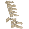
Figure 8
Figure 8. C6 sublaminar wire
Illustration of sublaminar wire shown passing the C6 pedicle screw. *This illustration is the authors' own creation.
Figure 9
Figure 9. C6-7 reduction via sequential tensioning
Illustration of C6–C7 reduction depicted after application of the rod followed by sequential wire tensioning. *This illustration is the authors' own creation.
Figure 10
Figure 10. C6-7 final reduction
Illustration of the final reduction and instrumentation of C6–C7 after sublaminar wire removal. *This illustration is the authors' own creation.
Figure 11
Figure 11. CT scan of the final C6-7 fracture reduction
(A) Sagittal spine CT demonstrating near total anatomical reduction of C6–C7 fracture dislocation; (B) axial spine CT of the corresponding level of C6–C7 vertebral body.

References

    1. Management of sub-axial cervical spine injuries. Zaveri G, Das G. Indian J Orthop. 2017;51:633–652. - PMC - PubMed
    1. Subaxial cervical spine trauma: evaluation and surgical decision-making. Joaquim AF, Patel AA. Global Spine J. 2014;4:63–70. - PMC - PubMed
    1. Sub-axial cervical facet dislocation: a review of current concepts. Mubark I, Abouelela A, Hassan M, Genena A, Ashwood N. Cureus. 2021;13:0. - PMC - PubMed
    1. Incidence rates and populations at risk for spinal cord injury: a regional study. Burke DA, Linden RD, Zhang YP, Maiste AC, Shields CB. Spinal Cord. 2001;39:274–278. - PubMed
    1. A novel operative approach for the treatment of old distractive flexion injuries of subaxial cervical spine. Liu P, Zhao J, Liu F, Liu M, Fan W. Spine (Phila Pa 1976) 2008;33:1459–1464. - PubMed

Publication types

LinkOut - more resources