. 2023 Jun 14;10(2):173-183.
doi: 10.3390/dermatopathology10020025.

TRPS1: A Marker of Follicular Differentiation

Affiliations

TRPS1: A Marker of Follicular Differentiation

Kristin J Rybski et al. Dermatopathology (Basel). .

Abstract

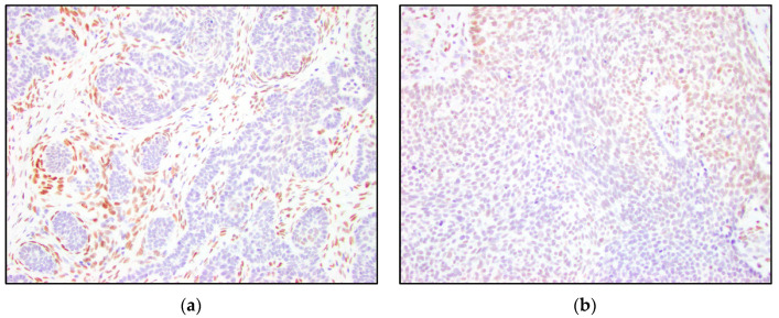
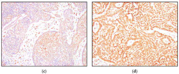
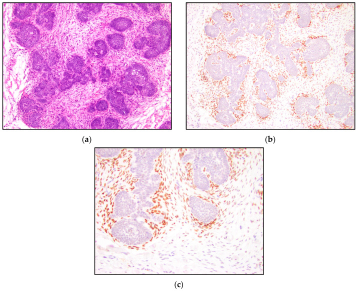
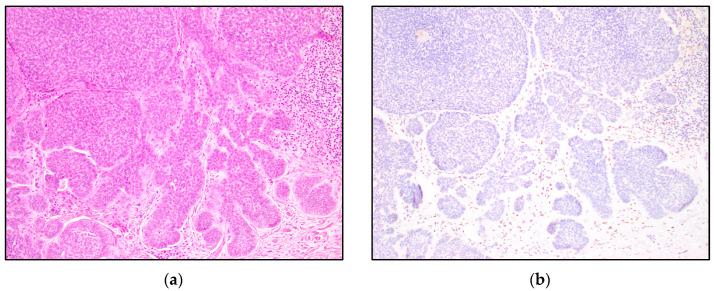
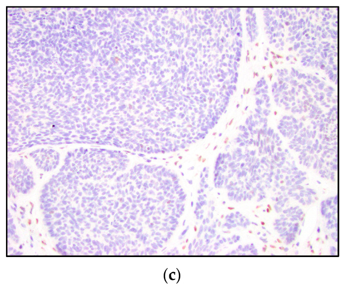
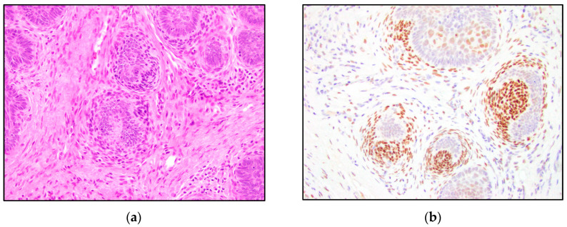
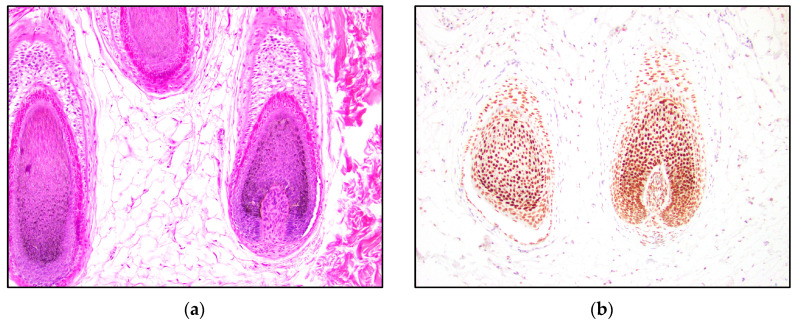
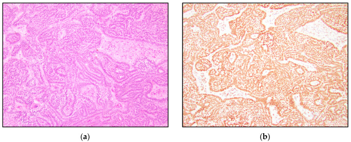
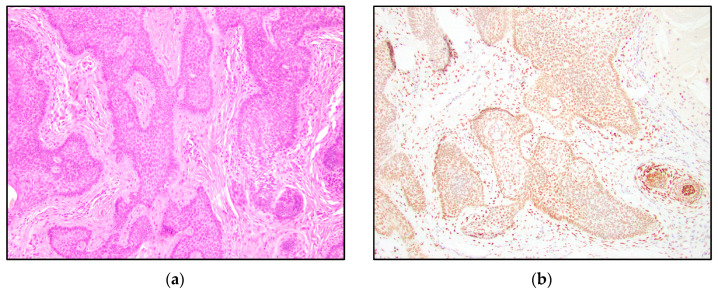
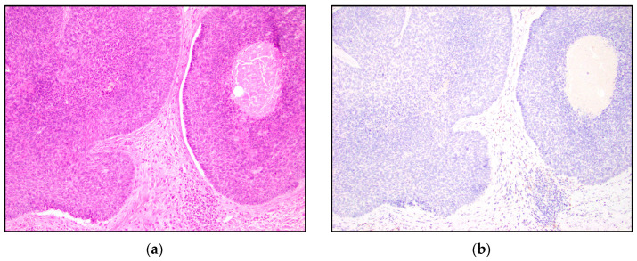
The trichorhinophalangeal syndrome type 1 (TRPS1) immunohistochemical (IHC) stain has increased in use in recent years as a marker for breast carcinomas. The TRPS1 gene is involved in various tissues, including the growth and differentiation of hair follicles. This article seeks to evaluate the IHC expression of TRPS1 in cutaneous neoplasms with follicular differentiation, such as trichoblastoma (TB), trichoepithelioma (TE), and basal cell carcinoma (BCC). IHC studies were performed on 13 TBs, 15 TEs, and 15 BCCs with an antibody against TRPS1. The study found a variable staining expression of TRPS1 in the tumor nests of TB, TE, and BCC. BCCs were distinct in that none of the BCCs demonstrated intermediate or high positivity, while TBs and TEs showed intermediate-to-high positivity in 5/13 (38%) and 3/15 (20%) of cases, respectively. We observed a distinct staining pattern among the mesenchymal cells of TB and TE. We found that TRPS1 highlighted perifollicular mesenchymal cells adjacent to the nests of TB and TE tumor cells. This staining pattern was absent in BCCs, where only scattered stromal cells were positive for TRPS1. Papillary mesenchymal bodies were also highlighted by TRPS1 in TB and TE. TRPS1 stained various parts of the normal hair follicle, including the nuclei of cells in the germinal matrix, outer root sheaths, and hair papillae. TRPS1 may be a useful IHC marker for follicular differentiation.

Keywords: TRPS1; basal cell carcinoma; follicular differentiation; hair follicle; trichoblastoma; trichoepithelioma.


Conflict of interest statement

The authors declare no conflict of interest.

Figures

Figure 1
Figure 1
Examples of TRPS1 immunoreactivity scores in the tumor nests. (a) Negative TRPS1 score in trichoepithelioma and positive staining of mesenchymal cells (200×); (b) low positive TRPS1 score in basal cell carcinoma (200×); (c) intermediate positive TRPS1 score in trichoepithelioma (200×); (d) high positive TRPS1 score in trichoblastoma (200×).
Figure 1
Figure 1
Examples of TRPS1 immunoreactivity scores in the tumor nests. (a) Negative TRPS1 score in trichoepithelioma and positive staining of mesenchymal cells (200×); (b) low positive TRPS1 score in basal cell carcinoma (200×); (c) intermediate positive TRPS1 score in trichoepithelioma (200×); (d) high positive TRPS1 score in trichoblastoma (200×).
Figure 2
Figure 2
TRPS1 IHC highlighting perifollicular mesenchymal cells in trichoblastoma. (a) Trichoblastoma (H&E, 100×); (b,c) TRPS1 strongly stained mesenchymal cells surrounding the tumor nests in trichoblastoma (100× and 200×).
Figure 3
Figure 3
TRPS1 IHC demonstrating scattered mesenchymal cells in basal cell carcinoma. (a) Basal cell carcinoma (H&E, 100×); (b,c) TRPS1 stains scattered mesenchymal cells in basal cell carcinoma (100× and 200×).
Figure 3
Figure 3
TRPS1 IHC demonstrating scattered mesenchymal cells in basal cell carcinoma. (a) Basal cell carcinoma (H&E, 100×); (b,c) TRPS1 stains scattered mesenchymal cells in basal cell carcinoma (100× and 200×).
Figure 4
Figure 4
TRPS1 IHC staining papillary mesenchymal bodies in trichoepithelioma. (a,b) Trichoepithelioma with papillary mesenchymal bodies highlighted by the TRPS1 stain (H&E and TRPS1, 200×).
Figure 5
Figure 5
TRPS1 IHC expression in normal hair follicles. (a) Normal hair follicle (H&E, 100×); (b) TRPS1 staining the root sheaths, germinative cells, matrix cells, and dermal papilla of a hair follicle (100×).
Figure 6
Figure 6
(a) Trichoblastoma (H&E, 100×). (b) High positive TRPS1 immunoreactivity in the trichoblastoma tumor nests (100×).
Figure 7
Figure 7
(a) Trichoepithelioma with a papillary mesenchymal body in the lower right corner (H&E, 100×). (b) High positive TRPS1 immunoreactivity in trichoepithelioma tumor nests and the papillary mesenchymal body (100×).
Figure 8
Figure 8
(a) Basal cell carcinoma with characteristic clefting (H&E, 100×). (b) Negative TRPS1 immunoreactivity in BCC tumor nests (100×).

References

    1. Ai D., Yao J., Yang F., Huo L., Chen H., Lu W., Soto L.M.S., Jiang M., Raso M.G., Wang S., et al. TRPS1: A highly sensitive and specific marker for breast carcinoma, especially for triple-negative breast cancer. Mod. Pathol. 2021;34:710–719. doi: 10.1038/s41379-020-00692-8. - DOI - PMC - PubMed
    1. Maas S.M., Shaw A.C., Bikker H., Lüdecke H.-J., van der Tuin K., Badura-Stronka M., Belligni E., Biamino E., Bonati M.T., Carvalho D.R., et al. Phenotype and genotype in 103 patients with tricho-rhino-phalangeal syndrome. Eur. J. Med. Genet. 2015;58:279–292. doi: 10.1016/j.ejmg.2015.03.002. - DOI - PubMed
    1. Momeni P., Glöckner G., Schmidt O., von Holtum D., Albrecht B., Gillessen-Kaesbach G., Hennekam R., Meinecke P., Zabel B., Rosenthal A., et al. Mutations in a new gene, encoding a zinc-finger protein, cause tricho-rhino-phalangeal syndrome type I. Nat. Genet. 2000;24:71–74. doi: 10.1038/71717. - DOI - PubMed
    1. Gai Z., Gui T., Muragaki Y. The function of TRPS1 in the development and differentiation of bone, kidney, and hair follicles. Histol. Histopathol. 2011;26:915–921. - PubMed
    1. Fantauzzo K.A., Bazzi H., Jahoda C.A., Christiano A.M. expression of the zinc-finger transcription factor Trps1 during hair follicle morphogenesis and cycling. Gene Expr. Patterns. 2008;8:51–57. doi: 10.1016/j.modgep.2007.10.006. - DOI - PubMed

LinkOut - more resources