

Raman Spectroscopy for Urea Breath Test
- PMID: 37366973
- PMCID: PMC10296114
- DOI: 10.3390/bios13060609
Raman Spectroscopy for Urea Breath Test
Abstract
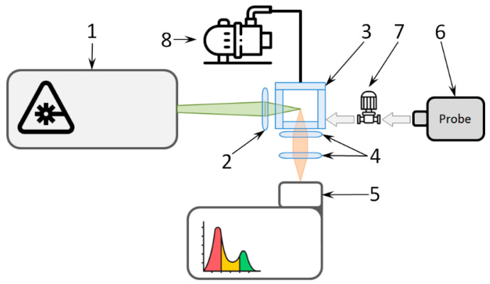
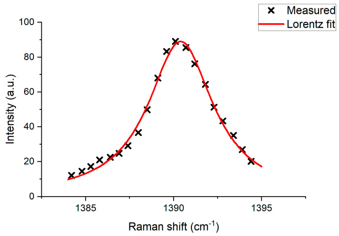
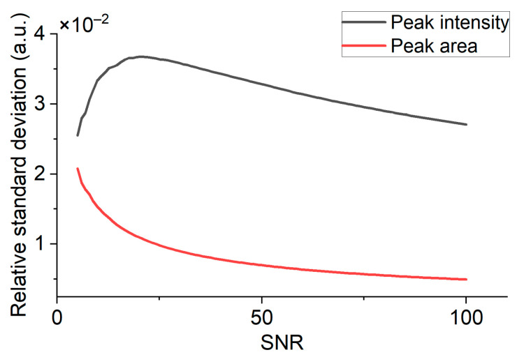
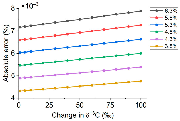
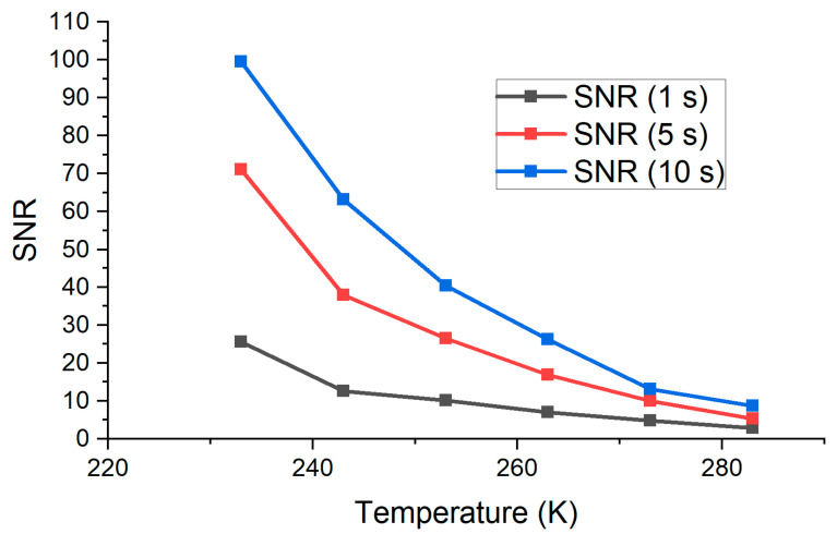
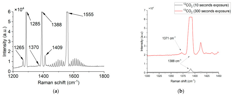
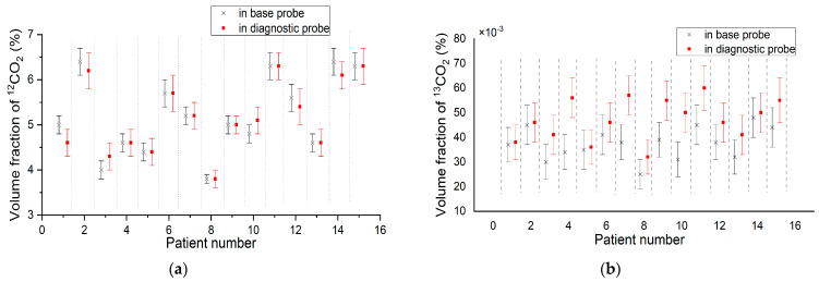
The urea breath test is a non-invasive diagnostic method for Helicobacter pylori infections, which relies on the change in the proportion of 13CO2 in exhaled air. Nondispersive infrared sensors are commonly used for the urea breath test in laboratory equipment, but Raman spectroscopy demonstrated potential for more accurate measurements. The accuracy of the Helicobacter pylori detection via the urea breath test using 13CO2 as a biomarker is affected by measurement errors, including equipment error and δ13C measurement uncertainty. We present a Raman scattering-based gas analyzer capable of δ13C measurements in exhaled air. The technical details of the various measurement conditions have been discussed. Standard gas samples were measured. 12CO2 and 13CO2 calibration coefficients were determined. The Raman spectrum of the exhaled air was measured and the δ13C change (in the process of the urea breath test) was calculated. The total error measured was 6% and does not exceed the limit of 10% that was analytically calculated.
Keywords: Helicobacter pylori; Raman spectroscopy; exhaled breath; urea breath test; δ13C.
Conflict of interest statement
The authors declare no conflict of interest.
Figures


Similar articles
-
Residual gas analyzer mass spectrometry for human breath analysis: a new tool for the non-invasive diagnosis of Helicobacter pylori infection.J Breath Res. 2014 Mar;8(1):016005. doi: 10.1088/1752-7155/8/1/016005. Epub 2014 Feb 24. J Breath Res. 2014. PMID: 24566134
-
Helicobacter pylori Breath Test via Mid-Infrared Sensor Technology.ACS Sens. 2025 Feb 28;10(2):1005-1010. doi: 10.1021/acssensors.4c02785. Epub 2025 Feb 8. ACS Sens. 2025. PMID: 39921651 Free PMC article.
-
Nondispersive infrared spectrometry for 13CO2/12CO2-measurements: a clinically feasible analyzer for stable isotope breath tests in gastroenterology.Z Gastroenterol. 1999 Jun;37(6):477-81. Z Gastroenterol. 1999. PMID: 10427653
-
The (13)C-urea breath test for non-invasive diagnosis of Helicobacter pylori infection: which procedure and which measuring equipment?Eur J Gastroenterol Hepatol. 2001 Jul;13(7):803-6. doi: 10.1097/00042737-200107000-00007. Eur J Gastroenterol Hepatol. 2001. PMID: 11474309 Review.
-
[C13 urea breath test in the diagnosis of Helicobacter pylori infection in the gastric mucosa. Validation of the method].Rev Esp Enferm Dig. 1996 Mar;88(3):202-8. Rev Esp Enferm Dig. 1996. PMID: 8645514 Review. Spanish.
Cited by
-
Reagentless Vis-NIR Spectroscopy Point-of-Care for Feline Total White Blood Cell Counts.Biosensors (Basel). 2024 Jan 19;14(1):0. doi: 10.3390/bios14010053. Biosensors (Basel). 2024. PMID: 38275306 Free PMC article.
References
-
- Chen Y., Zhang Y., Pan F., Liu J., Wang K., Zhang C., Gabriel A., Jesús M., Chen D., Cui D. Breath analysis based on surface-enhanced Raman scattering sensors distinguishes early and advanced gastric cancer patients from healthy persons. ACS Nano. 2016;10:8169–8179. doi: 10.1021/acsnano.6b01441. - DOI - PubMed
-
- Jaimes A.L., Durán C.M., Gualdrón O.E., Ionescu S.R. Stomach cancer detection through exhaled breath using biomarkers analysis. Chem. Eng. 2018;68:43–48.
MeSH terms
Substances
Grants and funding
LinkOut - more resources
Full Text Sources
Other Literature Sources
Medical
