Review
. 2023 Jun 4;13(6):617.
doi: 10.3390/bios13060617.

Aptameric Fluorescent Biosensors for Liver Cancer Diagnosis

Affiliations
Review

Aptameric Fluorescent Biosensors for Liver Cancer Diagnosis

Seonga Park et al. Biosensors (Basel). .

Abstract

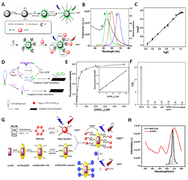
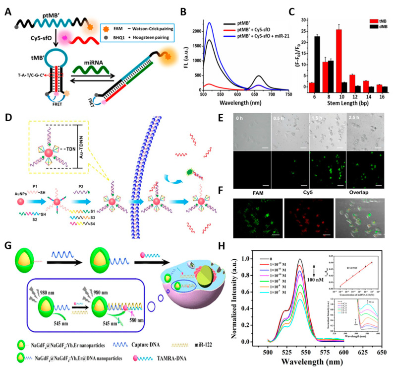
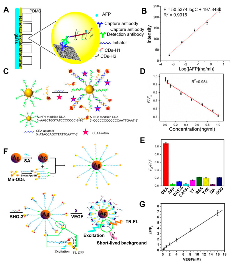
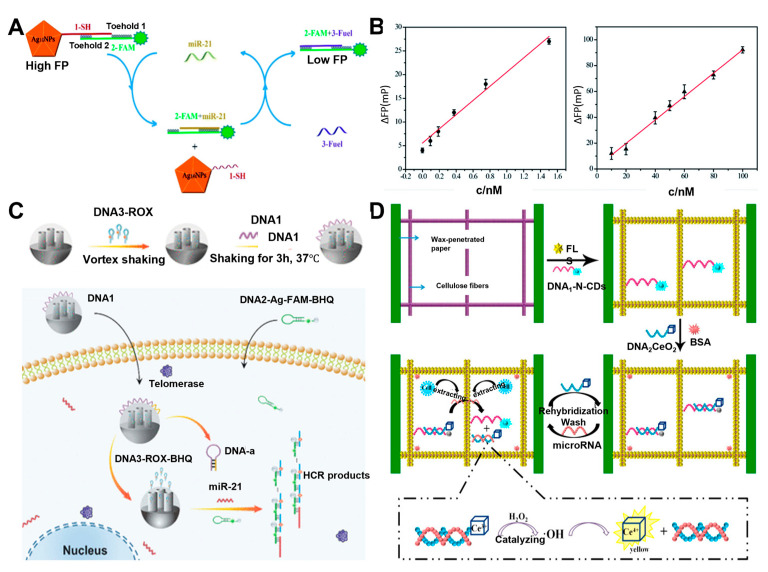
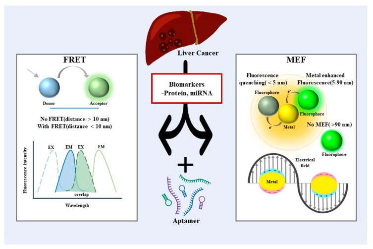
Liver cancer is a prevalent global health concern with a poor 5-year survival rate upon diagnosis. Current diagnostic techniques using the combination of ultrasound, CT scans, MRI, and biopsy have the limitation of detecting detectable liver cancer when the tumor has already progressed to a certain size, often leading to late-stage diagnoses and grim clinical treatment outcomes. To this end, there has been tremendous interest in developing highly sensitive and selective biosensors to analyze related cancer biomarkers in the early stage diagnosis and prescribe appropriate treatment options. Among the various approaches, aptamers are an ideal recognition element as they can specifically bind to target molecules with high affinity. Furthermore, using aptamers, in conjunction with fluorescent moieties, enables the development of highly sensitive biosensors by taking full advantage of structural and functional flexibility. This review will provide a summary and detailed discussion on recent aptamer-based fluorescence biosensors for liver cancer diagnosis. Specifically, the review focuses on two promising detection strategies: (i) Förster resonance energy transfer (FRET) and (ii) metal-enhanced fluorescence for detecting and characterizing protein and miRNA cancer biomarkers.

Keywords: Förster resonance energy transfer; aptamer; biosensors; liver cancer; metal-enhanced fluorescent.


Conflict of interest statement

The authors declare no conflict of interest.

Figures

Figure 2
Figure 2
(A) Illustration of a fluorescent sensor based on the FRET and has a sandwich structure involving QDs, AFP, and AuNPs. (B) Fluorescence spectroscopy to obtain ideal donor−acceptor pairs. (C) The linear detection range between the energy transfer efficiency and AFP concentrations. (D) Schematic illustration of the process for biomolecule detection. (E) The fluorescence intensity with the concentration of VEGF. The inset indicates the linear relationship. (F) Selectivity of VEGF with other biomolecules. (G) NIR−CDs−based fluorescence aptasensor for CEA detection. (H) The overlap between the emission spectrum of NIR−CDs and the absorption spectrum of AuNRs. (A−C): Reproduced with permission from [32], published by Talanta 2019. (D−F): Reproduced with permission from [37], published by Analyst 2015. (G,H): Reproduced with permission from [38], published by Analytica Chimica Acta 2019.
Figure 3
Figure 3
(A) Schematic illustration of the detection of miRNA using the tMB. (B) Fluorescence emission spectra of tMB formation and target addition. (C) The fluorescence recovery degrees according to different triplex or duplex lengths. (D) Structure and working principle of Au−TDNNs. (E) Uptake efficiency and dynamics of Au−TDNNs in the presence of miR−21 in live cells. Scale bars = 50 µm. (F) Strong FRET signal (red fluorescence) for miR−21 in HepG2 cells. Scale bars = 50 µm. (G) Illustration of the Synthesis and Workflow of UCNPs@DNA Nanoparticles for Sensitive Detection of miR−122. (H) Emission spectra of UCNPs@DNA nanoparticles with different concentrations of miR−122. The insets show the linear relationship (up) and the enlarged view of the emission at 580 nm (down). (AC): Reproduced with permission from [47], published by ACS Sensors 2018. (DF): Reproduced with permission from [48], published by Theranostics 2018. (G,H): Reproduced with permission from [51], published by ACS Applied Materials & Interfaces 2018.
Figure 4
Figure 4
(A) Schematic illustration of the operation to detect AFP with the Immuno−HCR and MEF of Carbon Dots. (B) Linear plot for fluorescence corresponding to concentrations of AFP. (C) Schematic illustration of the MEF strategy based on the two different types of nanomaterials for assaying CEA. (D) Linear plot of F/F0 versus concentrations of CEA from 0.01 ng/mL to 1 ng/mL. (E) Selectivity of CEA (0.8 ng/mL) with other related proteins. (F) Schematic illustration of the fabrication procedures of the silver nanoparticle−enhanced TR−FL sensor based on the Mn−doped ZnS QDs and the detection mechanism for VEGF165. (G) Linear plot of ΔF/F0 to VEGF165 concentration and TR-FL sensor specificity for VEFG165 (A,B): Reproduced with permission from [56], published by ACS Appl. Mater. Interfaces 2017. (CE): Reproduced with permission from [58], published by ScienceDirect Biosensors and Bioelectronics 2015. (F,G): Reproduced with permission from [59], published by ScienceDirect Biosensors and Bioelectronics 2015.
Figure 5
Figure 5
(A) Schematic illustration of biosensor based on Cyclic strand displacement reaction for detecting miR−21. (B) Linear plot of the fluorescence polarization change (ΔFP) corresponding to the concentrations of miRNA−21 with and without cyclic strand displacement reaction. (C) Schematic of the structure and operation of FOMN−based dual−signal logic. (D) Schematic illustration of the fabrication procedures and detection mechanism. (A,B): Reproduced with permission from [63], published by RSC Advances 2020. (C): Reproduced with permission from [64], published by ACS Appl. Mater. Interfaces 2021. (D): Reproduced with permission from [67], published by ScienceDirect Biosensors and Bioelectronics 2017.
Figure 1
Figure 1
Schematic illustration of aptameric fluorescent biosensors for liver cancer diagnosis.

Similar articles

Cited by

References

    1. Bray F., Laversanne M., Weiderpass E., Soerjomataram I. The ever-increasing importance of cancer as a leading cause of premature death worldwide. Cancer. 2021;127:3029–3030. doi: 10.1002/cncr.33587. - DOI - PubMed
    1. Sung H., Ferlay J., Siegel R.L., Laversanne M., Soerjomataram I., Jemal A., Bray F. Global Cancer Statistics 2020: GLOBOCAN Estimates of Incidence and Mortality Worldwide for 36 Cancers in 185 Countries. CA Cancer J. Clin. 2021;71:209–249. doi: 10.3322/caac.21660. - DOI - PubMed
    1. Ferlay J., Colombet M., Soerjomataram I., Parkin D.M., Pineros M., Znaor A., Bray F. Cancer statistics for the year 2020: An overview. Int. J. Cancer. 2021;149:778–789. doi: 10.1002/ijc.33588. - DOI - PubMed
    1. Anwanwan D., Singh S.K., Singh S., Saikam V., Singh R. Challenges in liver cancer and possible treatment approaches. Biochim. Biophys. Acta Rev. Cancer. 2020;1873:188314. doi: 10.1016/j.bbcan.2019.188314. - DOI - PMC - PubMed
    1. Zong J., Fan Z., Zhang Y. Serum Tumor Markers for Early Diagnosis of Primary Hepatocellular Carcinoma. J. Hepatocell. Carcinoma. 2020;7:413–422. doi: 10.2147/JHC.S272762. - DOI - PMC - PubMed

MeSH terms