. 2023 May 29;45(6):4687-4700.
doi: 10.3390/cimb45060298.

Acceleration of the Deamination of Cytosine through Photo-Crosslinking

Affiliations

Acceleration of the Deamination of Cytosine through Photo-Crosslinking

Siddhant Sethi et al. Curr Issues Mol Biol. .

Abstract

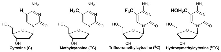
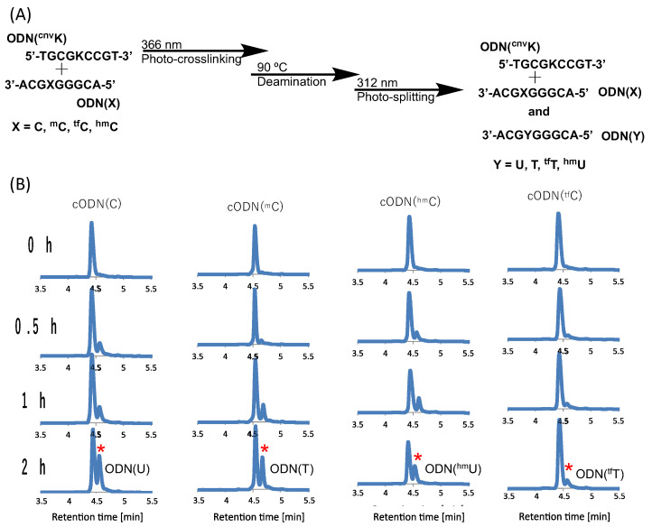
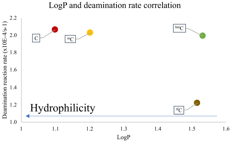
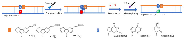
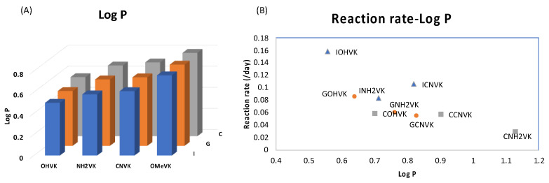
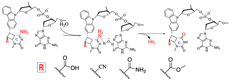
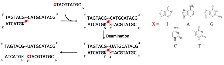
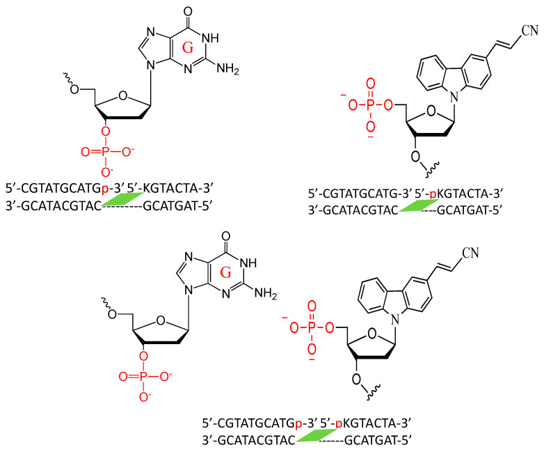
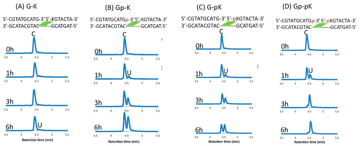
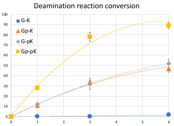
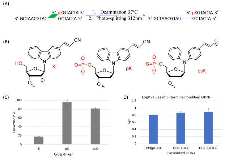
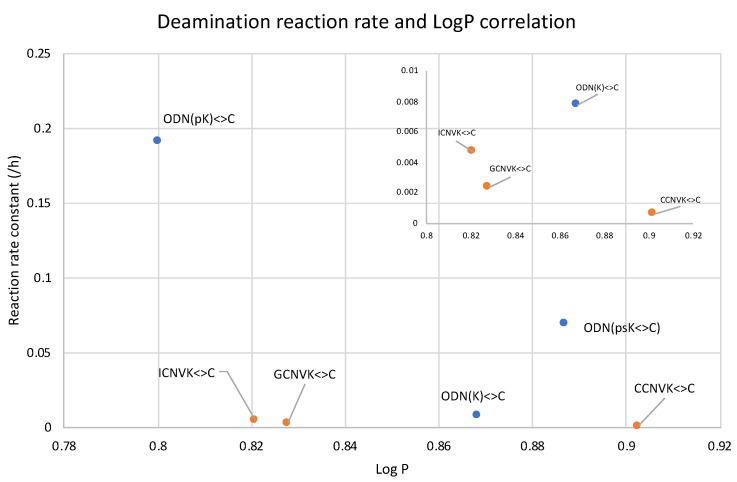
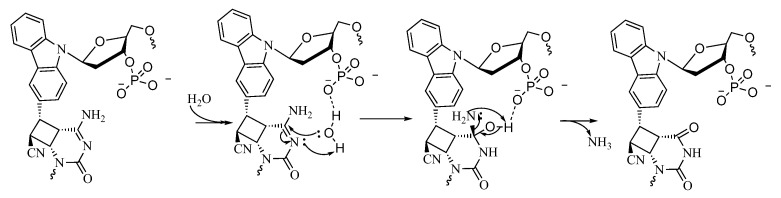
Herein, we report the major factor for deamination reaction rate acceleration, i.e., hydrophilicity, by using various 5-substituted target cytosines and by carrying out deamination at high temperatures. Through substitution of the groups at the 5'-position of the cytosine, the effect of hydrophilicity was understood. It was then used to compare the various modifications of the photo-cross-linkable moiety as well as the effect of the counter base of the cytosine to edit both DNA and RNA. Furthermore, we were able to achieve cytosine deamination at 37 °C with a half-life in the order of a few hours.

Keywords: DNA manipulation; RNA editing; cytosine deamination; nucleobase editing; photo-crosslinking.


Conflict of interest statement

The authors declare no conflict of interest.

Figures

Figure 1
Figure 1
Structure of the cytosine derivatives.
Figure 2
Figure 2
(A) Scheme of the photo-crosslink-assisted cytosine deamination reaction using cytosine derivatives. (B) UPLC analysis of the deamination reaction of the photo-cross-linked duplex consisting of ODN(K) and cODN(C) consisting of cytosine derivatives. [ODN(K<>C)] = 5 μM in 50 mM Na-cacodylate buffer (pH 7.4) containing 100 mM NaCl. incubated at 37 °C; photo splitting was performed with a transilluminator (312 nm) at 37 °C. The peak marked with an asterisk (*) indicates the newly formed product as depicted on each plot.
Figure 3
Figure 3
Dependence of LogP and the reaction rate constant of the deamination reaction at 90 °C.
Figure 4
Figure 4
Scheme of deamination of cytosine with different counter base and photo-crosslinker incorporated in the ODN.
Figure 5
Figure 5
(A) Bar graph representing the LogP of each combination of ODN containing a counter base (I, G, or C) and a photo-crosslinker (OHVK, OMeVK, CNVK, or NH2VK). The detailed data are provided in Table S5. Lower LogP values represent higher hydrophilicity. (B) Correlation of LogP and the reaction rate constant of the deamination reaction at 37 °C.
Figure 6
Figure 6
Proposed mechanism of the deamination of cytosine cross-linked to the cross-linker in the complementary strand.
Figure 7
Figure 7
Scheme of deamination of the cytosine with disjointed complimentary ODNs. Here, K is CNVK and X is the counter base of the target cytosine.
Figure 8
Figure 8
Conversion of cytosine to uracil after the photo-crosslink-assisted deamination reaction with ODN(K) and ODN(X/Xp).
Figure 9
Figure 9
Sequence and structure of the disjointed ODNs with phosphate-modified guanine and CNVK.
Figure 10
Figure 10
UPLC analysis of the deamination reaction of the photo-cross-linked duplex consisting of ODN(K/pK), ODN(X/Xp), and ODN(tC). [ODN(G/K<>C)] = 5 μM in 50 mM Na-cacodylate buffer (pH 7.4) containing 100 mM NaCl. incubated at 37 °C; photo splitting was performed with a transilluminator (312 nm) at 37 °C.
Figure 11
Figure 11
Conversion of cytosine to uracil after the photo-crosslink-assisted deamination reaction with ODN(K/pK) and ODN(X/Xp).
Figure 12
Figure 12
(A) Deamination reaction scheme with 5′-terminal modified ODNs. (B) Structure of the various 5′-terminal modifications. (C) Conversion ratio of cytosine to uracil using the different 5′-terminal modifications. (D) LogP values of the 5′-terminal modified ODNs.
Figure 13
Figure 13
Compiled LogP values of the counterbase–crosslinker variation compared with 5′phosphate modification.
Figure 14
Figure 14
Proposed mechanism of the deamination of the cytosine cross-linked to the cross-linker in the complementary strand with a free phosphate group.

Similar articles

References

    1. Ikebe S., Tanaka M., Ozawa T. Point mutations of mitochondrial genome in Parkinson’s disease. Mol. Brain Res. 1995;28:281–295. doi: 10.1016/0169-328X(94)00209-W. - DOI - PubMed
    1. Sakuraba H., Oshima A., Fukuhara Y., Shimmoto, Nagao Y., Bishop D.F., Desnick R.J., Suzuki Y. Identification of point mutations in the alpha-galactosidase A gene in classical and atypical hemizygotes with Fabry disease. Am. J. Hum. Genet. 1990;47:784–789. - PMC - PubMed
    1. Li J., Uversky V.N., Fink A.L. Effect of familial Parkinson’s disease point mutations A30P and A53T on the structural properties, aggregation, and fibrillation of human alpha-synuclein. Biochemistry. 2001;40:11604–11613. doi: 10.1021/bi010616g. - DOI - PubMed
    1. De Vries D.D., van Engelen B.G., Gabreëls F.J., Ruitenbeek W., van Oost B.A. A second missense mutation in the mitochondrial ATPase 6 gene in Leigh’s syndrome. Ann. Neurol. 1993;34:410–412. doi: 10.1002/ana.410340319. - DOI - PubMed
    1. Lake N.J., Compton A.G., Rahman, Thorburn D.R. Leigh syndrome: One disorder, more than 75 monogenic causes. Ann. Neurol. 2016;79:190–203. doi: 10.1002/ana.24551. - DOI - PubMed

LinkOut - more resources