. 2023 May 31;45(6):4778-4795.
doi: 10.3390/cimb45060304.

Large-Scale Production of Anti-RNase A VHH Expressed in pyrG Auxotrophic Aspergillus oryzae

Affiliations

Large-Scale Production of Anti-RNase A VHH Expressed in pyrG Auxotrophic Aspergillus oryzae

Elif Karaman et al. Curr Issues Mol Biol. .

Abstract

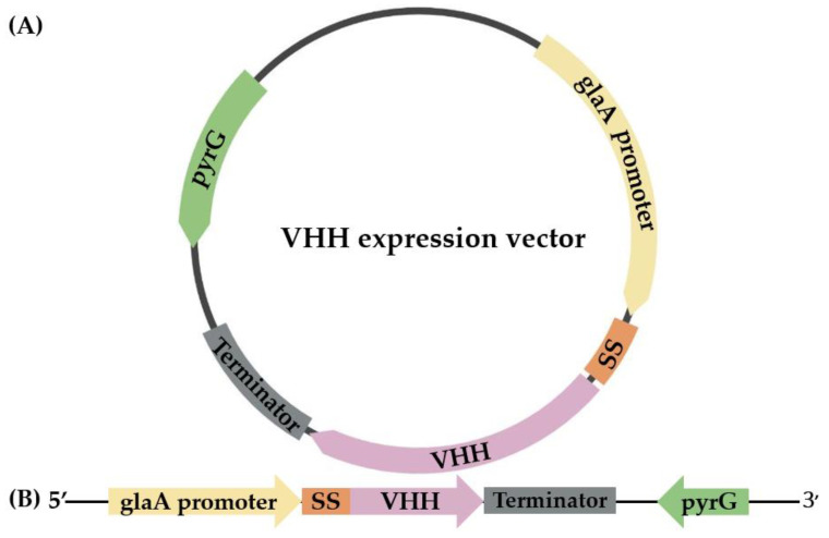
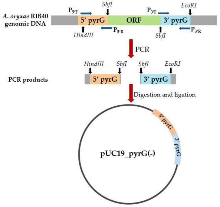
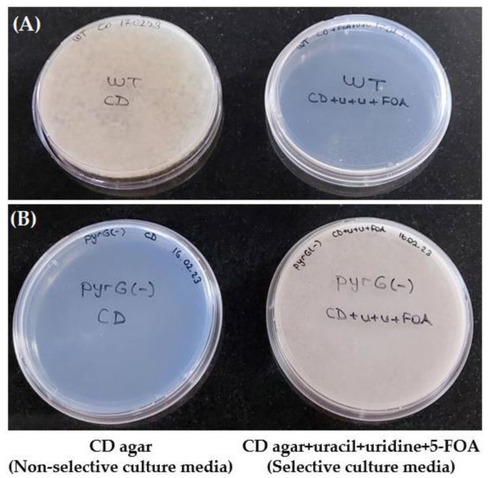
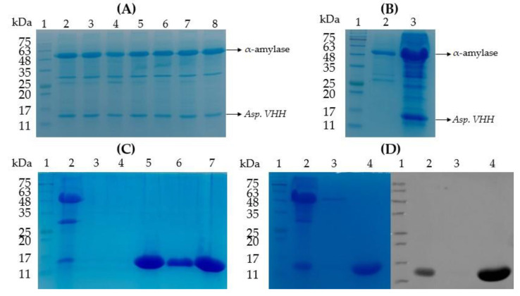
Nanobodies, also referred to as VHH antibodies, are the smallest fragments of naturally produced camelid antibodies and are ideal affinity reagents due to their remarkable properties. They are considered an alternative to monoclonal antibodies (mAbs) with potential utility in imaging, diagnostic, and other biotechnological applications given the difficulties associated with mAb expression. Aspergillus oryzae (A. oryzae) is a potential system for the large-scale expression and production of functional VHH antibodies that can be used to meet the demand for affinity reagents. In this study, anti-RNase A VHH was expressed under the control of the glucoamylase promoter in pyrG auxotrophic A. oryzae grown in a fermenter. The feature of pyrG auxotrophy, selected for the construction of a stable and efficient platform, was established using homologous recombination. Pull-down assay, size exclusion chromatography, and surface plasmon resonance were used to confirm the binding specificity of anti-RNase A VHH to RNase A. The affinity of anti-RNase A VHH was nearly 18.3-fold higher (1.9 nM) when expressed in pyrG auxotrophic A. oryzae rather than in Escherichia coli. This demonstrates that pyrG auxotrophic A. oryzae is a practical, industrially scalable, and promising biotechnological platform for the large-scale production of functional VHH antibodies with high binding activity.

Keywords: Aspergillus oryzae; VHH; affinity reagent; nanobodies; pyrG; single domain antibodies; surface plasmon resonance.


Conflict of interest statement

The authors declare no conflict of interest.

Figures

Figure 1
Figure 1
A schematic representation of circular (A) and linear (B) VHH expression vectors comprising the glucoamylase (glaA) promoter, the glucoamylase signal sequence (SS), the gene encoding anti-RNase A VHH (VHH), the terminator, and the pyrG gene.
Figure 2
Figure 2
The construction of the pyrG deletion vector (pUC19_pyrG(-)) is schematically depicted. The pyrG deletion vector was created by combining the 1.3 kb upstream fragment (5′ pyrG) and 1.2 kb downstream fragment (3′ pyrG) of A. oryzae RIB40 genomic DNA. PCR was used to amplify the 5′ and 3′ pyrG sequences without the pyrG open reading frame (ORF). P5′F–P5′R and P3′F–P3′R are primer pairs for 5′ pyrG and 3′ pyrG, respectively. To obtain pUC19_pyrG(-), PCR products were digested with HindIII, SbfI, and EcoRI restriction enzymes and ligated into the pUC19 vector backbone.
Figure 3
Figure 3
An illustration of the growth potential of pyrG(-) and wild-type (WT) A. oryzae on selective and non-selective culture media. CD agar culture medium plates are non-selective. CD agar plates of culture medium mixed with uracil, uridine, and 5-FOA are selective. (A) Wild-type A. oryzae can grow on non-selective media (left) but not on selective media in the presence of 5-FOA (right). (B) pyrG(-) A. oryzae cannot grow on a non-selective media lacking uracil and uridine (left) but can grow on selective media in the presence of uracil, uridine, and 5-FOA (right). (U+u+FOA: uracil, uridine and 5-fluoroorotic acid).
Figure 4
Figure 4
Sodium dodecyl sulfate polyacrylamide gel electrophoresis (SDS-PAGE) and Western blot studies of 8xHis-tagged Asp. VHH (~13 kDa) expressed in pyrG(-) A. oryzae. (A) SDS-PAGE profile of the small-scale protein expression of multiple transformed colonies after A. oryzae transformation. Lane 1: Gangnam-Stain Protein Ladder. Lanes 2–8: Culture medium samples from transformed colonies expressing Asp. VHH. (B) SDS-PAGE profile of the mock transfection control and a recombinant colony expressing Asp. VHH. Lane 1: Gangnam-Stain Protein Ladder. Lane 2: Culture medium of the mock transfection control. Lane 3: Culture medium of the recombinant colony expressing Asp. VHH. (C) SDS-PAGE analysis of purified Asp. VHH from large-scale production in a 6-liter fermenter by IMAC. Lane 1: Gangnam-Stain Protein Ladder. Lane 2: Concentrated culture medium sample of the recombinant colony expressing Asp. VHH. Lanes 3 and 4: The samples obtained from the IMAC wash steps. Lanes 5–7: The eluted sample containing Asp. VHH collected from the IMAC column. (D) Verification of purified Asp. VHH by SDS-PAGE (left) and Western blot (right) analyses, where for both the gel and blot, lane 1 is the Gangnam-Stain Protein Ladder, lane 2 is the culture medium of the recombinant colony expressing Asp. VHH, lane 3 is the sample obtained from IMAC wash step, lane 4 is the eluted sample containing Asp. VHH collected from the IMAC column.
Figure 5
Figure 5
Chromatogram of size exclusion chromatography using a Superdex 200 Increase 10/300 GL column. The peak represents pure Asp. VHH.
Figure 6
Figure 6
Pull-down assay to investigate the specificity of Asp. VHH against RNase A. The control resin (Cont.) was incubated with only RNase A. The experimental resin (Exp.) was co-incubated with Asp. VHH and RNase A. Lane 1: Flow-through sample containing only Asp. VHH (experimental resin). Lane 2: Wash sample obtained from the experimental resin after incubation with Asp. VHH. Lane 3: Gangnam-Stain Protein Ladder. Lane 4: Flow-through sample collected after co-incubation of Asp. VHH and RNase A (experimental resin). Lane 5: Flow-through sample containing only RNase A (control resin). Lane 6: Wash sample obtained from the experimental resin after RNase A incubation. Lane 7: Wash sample obtained from the control resin after RNase A incubation. Lane 8: Elution sample collected from the experimental resin containing RNase A and Asp. VHH. Lane 9: Elution sample collected from the control resin. Exp: experimental resin, Cont: control resin, FT: Flow-through sample, W: wash sample, and Elu: elution sample.
Figure 7
Figure 7
Size-exclusion chromatographic profiles on the Superdex 200 Increase 10/300 GL column of Asp. VHH, RNase A, and the mixture of Asp. VHH and RNase A. (A) Asp. VHH was applied to the size exclusion column; the green arrow denotes the peak of Asp. VHH. (B) RNase A was applied to the size exclusion column; the orange arrow denotes the peak of RNase A. (C) After Asp. VHH and RNase A were incubated together, the mixture was applied to the size exclusion column. The orange arrow represents the peak of RNase A, while the red arrow represents the complex of Asp. VHH and RNase A.
Figure 8
Figure 8
Sensorgrams display measured curves, whereas black lines depict fitted curves. The red curve represents the lowest RNase A concentration (2 nM), the green line indicates mid-concentration of RNase A (10 nM), whereas the blue curve represents the highest RNase A concentration (50 nM). A blank cycle was subtracted from each curve, and the binding activity of RNase A was measured. Asp. VHH was captured on the surface of the flow cell. The kinetics demonstrates an interaction between increasing concentrations of RNase A and Asp. VHH.

Similar articles

Cited by

References

    1. Hamers-Casterman C., Atarhouch T., Muyldermans S., Robinson G., Hamers C., Bajyana Songa E., Bendahman N., Hammers R. Naturally occurring antibodies devoid of light chains. Nature. 1993;363:446–448. doi: 10.1038/363446a0. - DOI - PubMed
    1. Vincke C., Muyldermans S. Introduction to heavy chain antibodies and derived Nanobodies. In: Saerens D., Muyldermans S., editors. Single Domain Antibodies. Methods in Molecular Biology. Volume 911. Humana Press; New York, NY, USA: 2012. pp. 15–26. - DOI - PubMed
    1. Nguyen V.K., Hamers R., Wyns L., Muyldermans S. Loss of splice consensus signal is responsible for the removal of the entire CH1 domain of the functional camel IGG2A heavy-chain antibodies. Mol. Immunol. 1999;36:515–524. doi: 10.1016/S0161-5890(99)00067-X. - DOI - PubMed
    1. Govaert J., Pellis M., Deschacht N., Vincke C., Conrath K., Muyldermans S., Saerens D. Dual beneficial effect of interloop disulfide bond for single domain antibody fragments. J. Biol. Chem. 2012;287:1970–1979. doi: 10.1074/jbc.M111.242818. - DOI - PMC - PubMed
    1. Perruchini C., Pecorari F., Bourgeois J.P., Duyckaerts C., Rougeon F., Lafaye P. Llama VHH antibody fragments against GFAP: Better diffusion in fixed tissues than classical monoclonal antibodies. Acta Neuropathol. 2009;118:685–695. doi: 10.1007/s00401-009-0572-6. - DOI - PubMed

LinkOut - more resources